Dowling–Degos disease - Rjme.ro
Dowling–Degos disease - Rjme.ro
Dowling–Degos disease - Rjme.ro
Create successful ePaper yourself
Turn your PDF publications into a flip-book with our unique Google optimized e-Paper software.
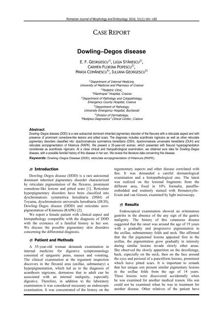
<st<strong>ro</strong>ng>Dowling–Degos</st<strong>ro</strong>ng> <st<strong>ro</strong>ng>disease</st<strong>ro</strong>ng> 183Figure 7 – Perivascular lympho-histiocytic infiltratewithin the superficial derma (HE stain, ×200). Discussion<st<strong>ro</strong>ng>Dowling–Degos</st<strong>ro</strong>ng> <st<strong>ro</strong>ng>disease</st<strong>ro</strong>ng> (DDD) is an autosomaldominant genodermatosis characterized by reticulatepigmentation of the flexures. This rare genodermatosiswas first described by Dowling GB and Freudenthal Win 1938 as a benign form of acanthosis nigricans [3],subsequent, she was termed “dermatose reticulée desplis” by Degos R and Ossipowski B in 1954 [4]. DDDwas first characterized by Wilson-Jones and Grice in1978 in their description of 10 patients with the disorderand recognized as a distinct entity [5]. It is an autosomaldominant inherited pigmentary disorder [6] usually ofadult onset, in individuals before they are aged 24 years,but may occur in childhood [1]. A Chinese newbornwith reticulate pigmented anomaly of the flexures wasrecently described [7].The <st<strong>ro</strong>ng>disease</st<strong>ro</strong>ng> affects both sexes, although, a femalepredominance has been noted in some surveys [8].DDD is often familial and appears to be inherited inan autosomal dominant manner [9]. Recently, a genelocus believed responsible in one Chinese patient wasmapped to 17p 13.3 [10] and a genome-wide linkageanalysis of two German families describe localization ofthe first DDD locus on ch<strong>ro</strong>mosome 12q/8/A [11]. Thisregion includes the keratin gene cluster, which wasscreened for mutations. Loss of function mutations wereidentified in the keratin 5 gene (KRT 5) in all affectedfamily members and in six unrelated patients withDDD. Keratin 5 is a component of the intermediate filament(IF) cytoskeleton in the basal layer of the keratinocytes[11]. Dysfunction of the IF cytoskeleton causesaberrant distribution of melanomes degradation suggestinga delayed degradation of melanin granules [11].Thus, in a family with DDD, a hete<strong>ro</strong>zygous frameshiftmutation in the V1-domain of keratin 5 was identified[12]. These data confirm that haplo-insufficiencyfor K5 causes DDD and points to a p<strong>ro</strong>minent <strong>ro</strong>le forthe keratin intermediate filament cytoskeleton withinbasal keratinocytes in epidermal pigment biology.Clinical manifestations of DDD or reticulate pigmentedanomaly of the flexures (RPAF) are dominated byspotted and reticulate pigmentation of the flexures [5, 6]which may be associated with dark comedone-likelesions and pitted scars.Figure 8 – Perivascular lympho-histiocytic infiltratewithin the superficial derma (HE stain, ×200).The pigmentation is p<strong>ro</strong>gressive, symmetrical, oftenextensive and completely asymptomatic [1]. This mostcommonly affects the axillae, g<strong>ro</strong>ins, submammaryfolds and neck, but sometimes can spread to involve theface, chest, perineum, natal clefts and wrists [13, 14].The pigmentation consists of nume<strong>ro</strong>us small, discrete<strong>ro</strong>und to oval pigmented macules which resemblefreckles. It may be intense, with a b<strong>ro</strong>wnish black colorand sometimes steel blue or navy overtones. However,if the condition is less severe, it is stippled in shades ofb<strong>ro</strong>wn [15]. In many of these disorders, they are frecklelikeand angulated and tend to join at their margins toform a reticulate pattern. The confluence of lesionstoward the vault of the axillae and the centre ofgenitocrural folds was observed by Sandhu K et al. [1].If the patches are palpable, it is because of lichenificationthat p<strong>ro</strong>duces a glossy and at times somewhatwrinkled appearance.The pigmentation undergoes slow g<strong>ro</strong>wth over theyears and worsens in summer [5, 6, 14].Other common features includes; hyperkeratoticcomedone-like dark-b<strong>ro</strong>wn follicular papules on theback and/or neck; pitted scars most characteristicallyoccur a<strong>ro</strong>und the lateral margins of the mouth, but caninvolve other areas of the face, neck, axilla, thighs, etc[1]; pruritus on the affected areas. In addition, speckledmacules involving the dorsum of the hands, the p<strong>ro</strong>ximalnail folds, or the sc<strong>ro</strong>tum [15] may be seen. Fingernaildyst<strong>ro</strong>phy may be present, particularly finger-likefib<strong>ro</strong>ma [8, 16]. The finding of speckled macules on thesc<strong>ro</strong>tum is isolated and limited to the sc<strong>ro</strong>tal and penileskin [15]. This pigmented eruption on the male externalgenitalia is possibly a cutaneous marker of underlyingtesticular carcinoma. In female patients, speckledmacules may be found on the vulva [17, 18].Various affections have been associated with this<st<strong>ro</strong>ng>disease</st<strong>ro</strong>ng>. Thus, the association of DDD and hidradenitissuppurativa (HS) [19] single or associated with other<st<strong>ro</strong>ng>disease</st<strong>ro</strong>ng> as well as multiple keratoacanthomas [20],multiple epidermal cysts [21] and perianal squamouscell carcinoma [22], multiple seborrheic warts is wellreported. The association of pigmented eruption on themale external genital has been considered as a possiblemarker of underlying testicular carcinoma [15], althoughthe association is p<strong>ro</strong>bably fortuitous. Concerning HS,
184it is thought that they could be due to clinical manifestationof a single underlying defect in follicularkeratinization. Identically, for the same of the otherconditions, their coexistence in the same patient is likelyto reflect the same follicular anomaly. It is possibly thata single underlying defect of follicular p<strong>ro</strong>liferation mayaccount for the coexistence of these conditions.Histological, the affected skin shows elongatedepidermal rete ridges with thinning of the suprapapillaryepithelium and basilar hyperpigmentation in afiliform pattern. Perivascular lympho-histiocytic dermalinfiltration and dermal fib<strong>ro</strong>sis along elongated reteridges are observed [8]. Also, intra-epidermalkeratincysts may be remarked [16]. Occasionally, hamartomatousepidermal changes have been identified [16, 22].The downward elongation is composed of regularpigmented basaloid cells. An increased number ofmelanophages has been observed in, with no quantitativeincrease in the number of melanocytes [5].In a latest study, all pigmented cells in the basallayer were recognized by anti-PEP-1, anti-PEP-2,HMB-45 and NK1/beteb antibodies. The melanocyteswere localized in the basal layer and accounted for 10%of the total keratinocytes. Supranuclear “caps” of b<strong>ro</strong>wngranules were observed within most basal keratinocytesin the hyperpigmentation area. The melanocytes containedmany mitochondria, Golgi apparatus, and regularmelanosomes in all stages of maturation in theircytoplasm; melanosome-laden dendrites were readilydetected by transmission elect<strong>ro</strong>n mic<strong>ro</strong>scope. Melanosomesmainly of stages III and IV were evident withinkeratinocytes either distributed as scattered patterns orforming “caps” over the nucleus [23].Differential diagnoses include acanthosis nigricansand other human genetic pigmental <st<strong>ro</strong>ng>disease</st<strong>ro</strong>ng>, such asreticulate ac<strong>ro</strong>pigmentation of Kitamura (RAPK), dysch<strong>ro</strong>matosissymmetrica hereditaria (DSH), dysch<strong>ro</strong>matosisuniversalis hereditaria (DUH).In acanthosis nigricans, the plaques are velvety andthere may be skin tags but no dark comedone-likelesions. Histologically, there is papillomatosis in acanthosisnigricans and no follicular anomalies.The onset of RAPK is in the first two decades oflife. The location of macules is acral rather thanflexural. There are palmar and plantar pits and breaks inthe epidermal ridge pattern, which were not found inthis patient. The histological features of DDD andRAPK are however, very similar; RAPK lacking onlythe antler-like pattern of the epithelial p<strong>ro</strong>liferation.Several authors reported that <st<strong>ro</strong>ng>Dowling–Degos</st<strong>ro</strong>ng> <st<strong>ro</strong>ng>disease</st<strong>ro</strong>ng>and RAPK might be different clinical expressions of thesame <st<strong>ro</strong>ng>disease</st<strong>ro</strong>ng> [10].DSH is an autosomal dominant inheritancecharacterized by pinpoint, pea-sized, hyperpigmented,and hypopigmented macules limited largely to thedorsal aspects of the hand and feet [24].DDD are dominated by spotted and reticulatepigmentation of the flexures. In addition, the gene forDSH has previously been mapped to ch<strong>ro</strong>mosome1q11–1q21 [25], in which the RNA-specific adenosinedeaminase gene was identified as the <st<strong>ro</strong>ng>disease</st<strong>ro</strong>ng> gene forDSH [10, 24, 26].E. F. Georgescu et al.Dysch<strong>ro</strong>matosis universalis hereditaria is characterizedby pigmented flecks and spots over much of thebody other than that limited to the flexures of body [26].In addition, the gene for dysch<strong>ro</strong>matosis universalishereditaria has previously been mapped to ch<strong>ro</strong>mosome6q24.2–q25.2 [26].DDD exhibits a slow p<strong>ro</strong>gressive course. Treatmentof DDD is complicated, usually with poor results.Transient good results have been reported with the useof topical corticoste<strong>ro</strong>ids, depigmentating agents,topical adapalene and Er: YAG laser pulse energybetween 1000 and 1200 mJ, three consecutive passes[27, 28]. These treatments led to a good clinical resultand might be a successful strategy in DDD. ConclusionsThe clinical aspects and the histopathological examinationdiscovered in our patient are correlated with thediagnosis of DDD, with possible familial history in herson. Treatment of DDD is complicated and usually whichpoor results. It necessary ample studies on a nume<strong>ro</strong>uspatients with this rare <st<strong>ro</strong>ng>disease</st<strong>ro</strong>ng> for evaluation the efficacyand safety of the different therapeutically variants.References[1] SANDHU K, SARASWAT A, KANWAR AJ, <st<strong>ro</strong>ng>Dowling–Degos</st<strong>ro</strong>ng><st<strong>ro</strong>ng>disease</st<strong>ro</strong>ng> with dysch<strong>ro</strong>matosis universalis hereditaria-likepigmentation in a family, J Eur Acad Dermatol Venereol,2004, 18(6):702–704.[2] OISO N, TSURUTA D, OTA T, KOBAYASHI H, KAWADA A,Spotted and rippled reticulate hypermelanosis: a possiblevariant of <st<strong>ro</strong>ng>Dowling–Degos</st<strong>ro</strong>ng> <st<strong>ro</strong>ng>disease</st<strong>ro</strong>ng>, Br J Dermatol, 2007,156(1):196–198.[3] DOWLING GB, FREUDENTHAL W, Acanthosis nigricans, Br JDermatol, 1938, 50:467–471.[4] DEGOS R, OSSIPOWSKI B, Dermatose pigmentaire réticuléedes plis, Ann Dermatol Syphiligr, 1954, 81:147–151.[5] WILSON JE, GRICE K, Reticulate pigmented anomaly ofthe flexures: <st<strong>ro</strong>ng>Dowling–Degos</st<strong>ro</strong>ng>, a new genodermatosis, ArchDermatol, 1978, 114:1150–1157.[6] CROVATO F, NAZZARI G, REBORA A, <st<strong>ro</strong>ng>Dowling–Degos</st<strong>ro</strong>ng> <st<strong>ro</strong>ng>disease</st<strong>ro</strong>ng>(reticulate pigmented anomaly of the flexures) is anautosomal dominant condition, Br J Dermatol, 1983,108(4):473–476.[7] ZUO YG, HO MG, JIN HZ, WANG BX, Case report: a case ofnewborn onset reticulate pigmented anomaly of the flexuresin a Chinese female, J Eur Acad Dermatol Venereol, 2008,22(7):901–903.[8] KIM YC, DAVIS MD, SCHANBACHER CF, SU WP, <st<strong>ro</strong>ng>Dowling–Degos</st<strong>ro</strong>ng> <st<strong>ro</strong>ng>disease</st<strong>ro</strong>ng> (reticulate pigmented anomaly of theflexure): a clinical and histopathologic study of 6 cases,J Am Acad Dermatol, 1999, 40(3):462–467.[9] BROWN WG, Reticulate pigmented anomaly of the flexures.Case reports and genetic investigation, Arch Dermatol,1982, 118(7):490–493.[10] LI CR, XING QH, LI M, QIN W, YUE XZ, ZHANG XJ, MA HJ,WANG DG, FENG GY, ZHU WY, HE L, A gene locusresponsible for reticulate pigmented anomaly of the flexuresmaps to ch<strong>ro</strong>mosome 17p13.3, J Invest Dermatol, 2006,126(6):1297–1301.[11] BETZ RC, PLANKO L, EIGELSHOVEN S, HANNEKEN S,PASTERNACK SM, BUSSOW H, VAN DEN BOGAERT K,WENZEL J, BRAUN-FALCO M, RUTTEN A, ROGERS MA,RUZICKA T, NÖTHEN MM, MAGIN TM, KRUSE R, Loss-offunctionmutations in the keratin 5 gene lead to <st<strong>ro</strong>ng>Dowling–Degos</st<strong>ro</strong>ng> <st<strong>ro</strong>ng>disease</st<strong>ro</strong>ng>, Am J Hum Genet, 2006, 78(3):510–519.[12] LIAO H, ZHAO Y, BATY DU, MCGRATH JA, MELLERIO JE,MCLEAN WH, A hete<strong>ro</strong>zygous frameshift mutation in theV1 domain of keratin 5 in a family with <st<strong>ro</strong>ng>Dowling–Degos</st<strong>ro</strong>ng><st<strong>ro</strong>ng>disease</st<strong>ro</strong>ng>, J Invest Dermatol, 2007, 127(2):298–300.
<st<strong>ro</strong>ng>Dowling–Degos</st<strong>ro</strong>ng> <st<strong>ro</strong>ng>disease</st<strong>ro</strong>ng> 185[13] BARDACH HG, <st<strong>ro</strong>ng>Dowling–Degos</st<strong>ro</strong>ng> <st<strong>ro</strong>ng>disease</st<strong>ro</strong>ng> with involvement ofthe scalp, Hautartz, 1981, 32(4):182–186.[14] REBORA A, CROVATO F, The spectrum of <st<strong>ro</strong>ng>Dowling–Degos</st<strong>ro</strong>ng><st<strong>ro</strong>ng>disease</st<strong>ro</strong>ng>, Br J Dermatol, 1984, 110:627–630.[15] SCHWARTZ RA, BIRNKRANT AP, BURGESS GH,STOLL HL JR, YAQUB M, FOX MD, Reticulate pigmentedanomaly, Cutis, 1980, 26(4):380–381.[16] RUBIO C, MAYOR M, MARTÍN MA, GONZÁLEZ-BEATO MJ,CONTRERAS F, CASADO M, Atypical presentation of <st<strong>ro</strong>ng>Dowling–Degos</st<strong>ro</strong>ng> <st<strong>ro</strong>ng>disease</st<strong>ro</strong>ng>, J Eur Acad Dermatol Venereol, 2006,20(9):1162–1164.[17] O’GOSHI K, TERUI T, TAGAMI H, <st<strong>ro</strong>ng>Dowling–Degos</st<strong>ro</strong>ng> <st<strong>ro</strong>ng>disease</st<strong>ro</strong>ng>affecting the vulva, Acta Derm Venereol, 2001, 81(2):148.[18] MASSONE C, HOFMANN-WELLENHOF R, Dermoscopy of<st<strong>ro</strong>ng>Dowling–Degos</st<strong>ro</strong>ng> <st<strong>ro</strong>ng>disease</st<strong>ro</strong>ng> of the vulva, Arch Dermatol, 2008,144(3):417–418.[19] DIXIT R, GEORGE R, JACOB M, SUDARSANAM TD,DANDA D, <st<strong>ro</strong>ng>Dowling–Degos</st<strong>ro</strong>ng> <st<strong>ro</strong>ng>disease</st<strong>ro</strong>ng>, hidradenitis suppurativaand arthritis in mother and daughter, Clin Exp Dermatol,2006, 31(3):454–456.[20] FENSKE NA, GROOVER CE. LOBER CW, ESPINOZA CG,<st<strong>ro</strong>ng>Dowling–Degos</st<strong>ro</strong>ng> <st<strong>ro</strong>ng>disease</st<strong>ro</strong>ng>, hidradenitis suppurativa, and multiplekeratoacanthomas. A disorder that may be caused bya single underlying defect in pilosebaceous epithelial p<strong>ro</strong>liferation,J Am Acad Dermatol, 1991, 24(5 Pt 2):888–892.[21] LOO WJ, RYTINA E, TODD PM, Hidradenitis suppurativa,<st<strong>ro</strong>ng>Dowling–Degos</st<strong>ro</strong>ng> and multiple epidermal cysts: a new follicula<strong>ro</strong>cclusion triad, Clin Exp Dermatol, 2004, 29(6):622–624.[22] UJIHARA M, KAMAKURA T, IKEDA M, KODAMA H, <st<strong>ro</strong>ng>Dowling–Degos</st<strong>ro</strong>ng> <st<strong>ro</strong>ng>disease</st<strong>ro</strong>ng> associated with squamous cell carcinomaon the dappled pigmentation, Br J Dermatology, 2002,147(3):568–571.[23] ZHANG RZ, ZHU WY, A study of immunohistochemical andelect<strong>ro</strong>n mic<strong>ro</strong>scopic changes in <st<strong>ro</strong>ng>Dowling–Degos</st<strong>ro</strong>ng> <st<strong>ro</strong>ng>disease</st<strong>ro</strong>ng>,J Dermatol, 2005, 32(1):12–18.[24] ZHANG XY, HE PP, LI M, HE CD, YAN KL, CUI Y, YANG S,ZHANG KY, GAO M, CHEN JJ, LI CR, JIN L, CHEN HD,XU SJ, HUANG W, Seven novel mutations of ADAR genein Chinese families and sporadic patients with dysch<strong>ro</strong>matosissymmetrica hereditaria (DSH), Hum Mutat, 2004,23(6):629–630.[25] ZHANG XJ, GAO M, LI M, LI M, LI CR, CUI Y, HE PP, XU SJ,XIONG XY, WANG ZX, YUAN WT, YANG S, HUANG W,Identification of a locus for dysch<strong>ro</strong>matosis symmetricahereditaria at ch<strong>ro</strong>mosome 1q11–1q21, J Invest Dermatol,2003, 120(5):776–780.[26] MIYAMURA Y, SUZUKI T, KONO M, INAGAKI K, ITO S,SUZUKI N, TOMITA Y, Mutations of the RNA-specific adenosinedeaminase gene (DSRAD) are involved in dysch<strong>ro</strong>matosissymmetrica hereditaria, Am J Hum Genet, 2003,73(3):693–699.[27] ALTOMARE G, CAPELLA GL, FRACCHIOLLA C, FRIGERIO E,Effectiveness of topical adapalene in <st<strong>ro</strong>ng>Dowling–Degos</st<strong>ro</strong>ng><st<strong>ro</strong>ng>disease</st<strong>ro</strong>ng>, Dermatology, 1999, 198(2):176–177.[28] WENZEL G, PETROW W, TAPPE K, GERDSEN R,UERLICH WP, BIEBER T, Treatment of <st<strong>ro</strong>ng>Dowling–Degos</st<strong>ro</strong>ng><st<strong>ro</strong>ng>disease</st<strong>ro</strong>ng> with Er:YAG-laser: result after 2.5 years, DermatolSurg, 2003, 29(11):1161–1162.Corresponding authorEugen Florin Georgescu, P<strong>ro</strong>fessor, MD, PhD, Department of Internal Medicine, University of Medicineand Pharmacy of Craiova, 2–4 Petru Rareş Street, 200349 Craiova, Romania; Phone +40744–782 136,Fax +40251–420 896, e-mail: efg@usa.netReceived: June 15 th , 2009Accepted: January 5 th , 2010


