Copyright
©The Author(s) 2024.
World J Clin Cases. Jan 6, 2024; 12(1): 217-223
Published online Jan 6, 2024. doi: 10.12998/wjcc.v12.i1.217
Published online Jan 6, 2024. doi: 10.12998/wjcc.v12.i1.217
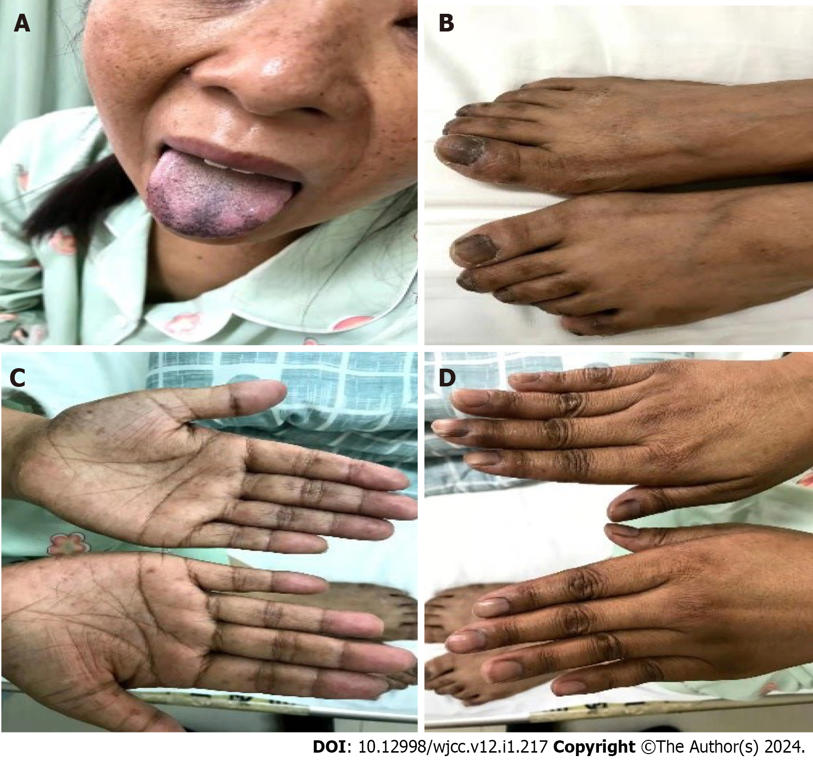
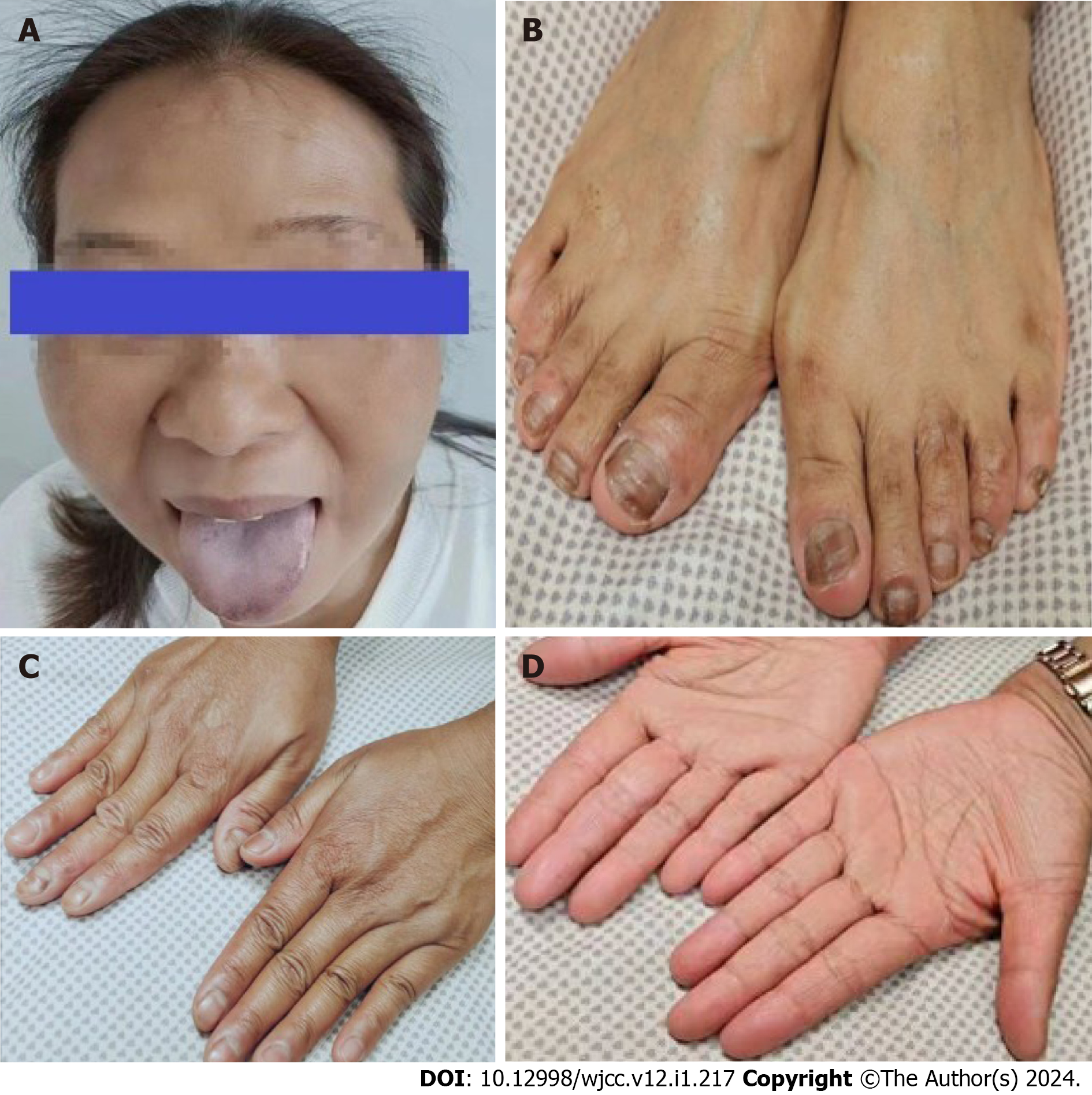
Figure 1 Multiple skin pigmentation.
A: Hyperpigmentation on the skin of face, mouth, tongue and mucous membranes of the lips; B-D: Hyperpigmentation on nails, hands and feet.
Figure 2 Histopathological analysis of the resected specimen.
A and B: Haematoxylin and eosin staining (× 40). Pathology of cutaneous tuberculosis; C: Skin ulcers on the back.
Figure 3 Computed tomography scan of pulmonary tuberculosis before treatment.
A and B: Upper field infection of both lungs; C: Computed tomography chest lung window coronal view shows bilateral lung infection.
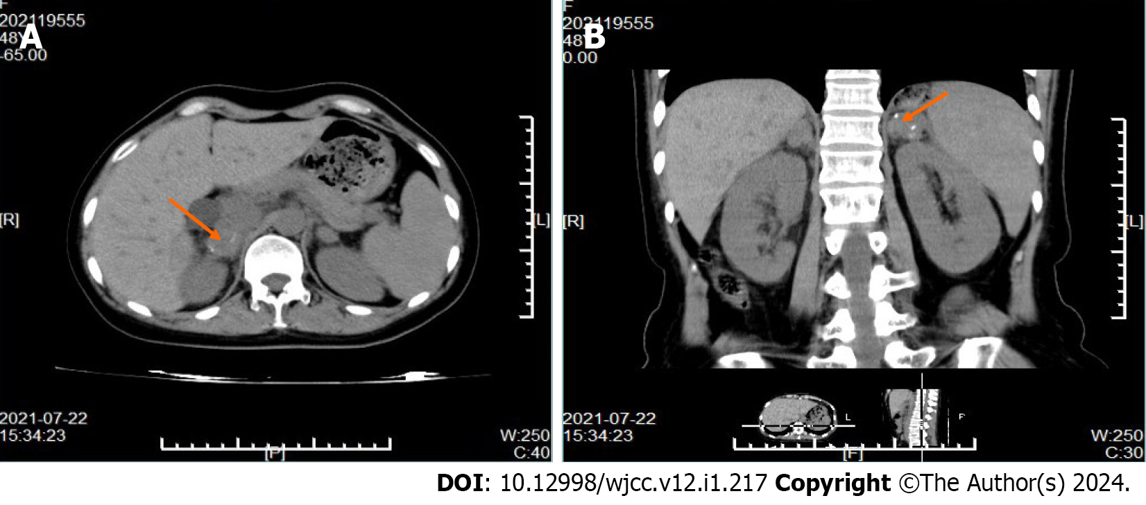
Figure 4 Adrenal computed tomography scan of adrenal enlargement and calcification (orange arrow).
A: Right adrenal gland enlargement and calcification at the orange arrow; B: Abdominal computed tomography coronal view shows enlargement and calcification of the right adrenal gland at the orange arrow.
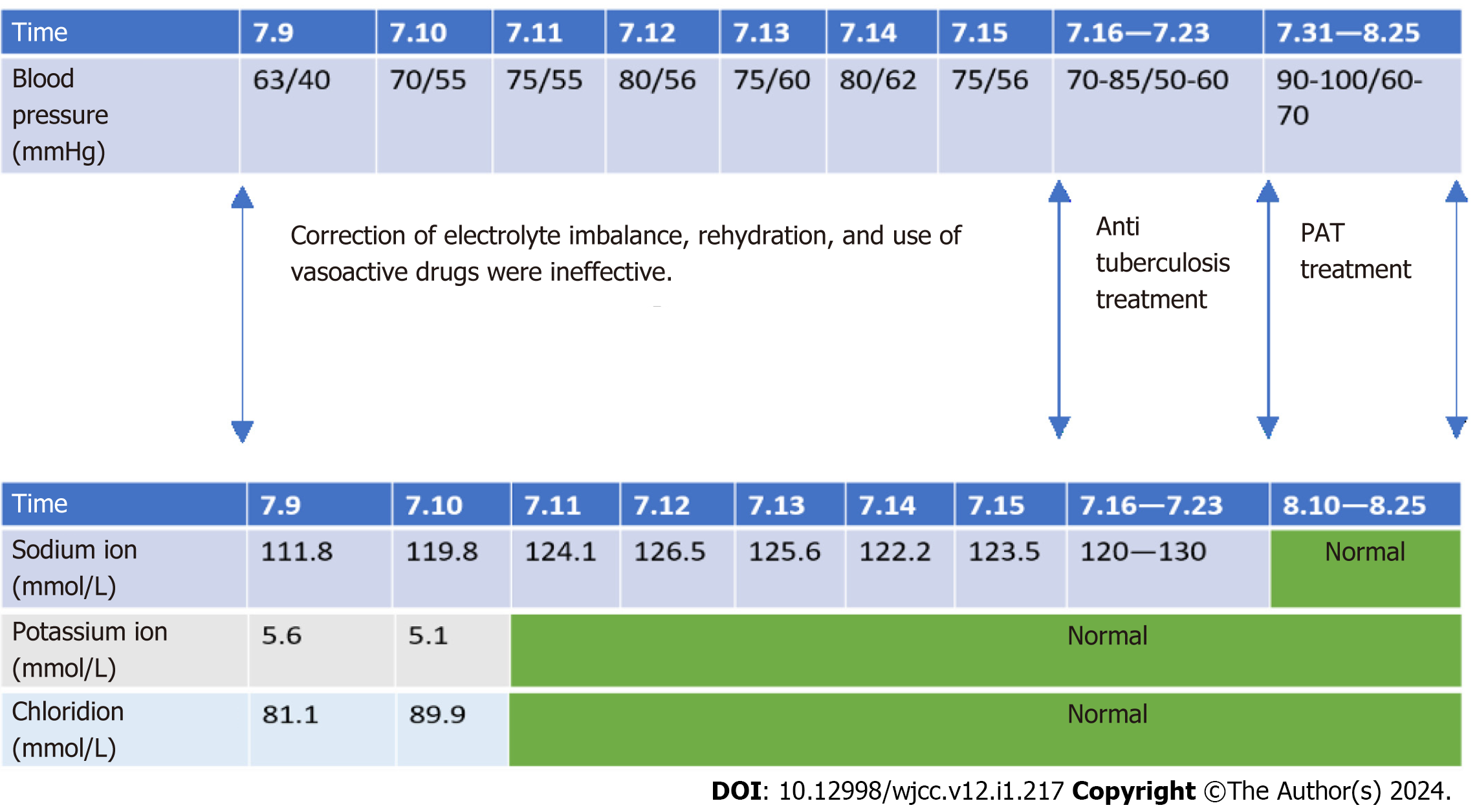
Figure 5 Time line table of the patient.
PAT: Prednisone.
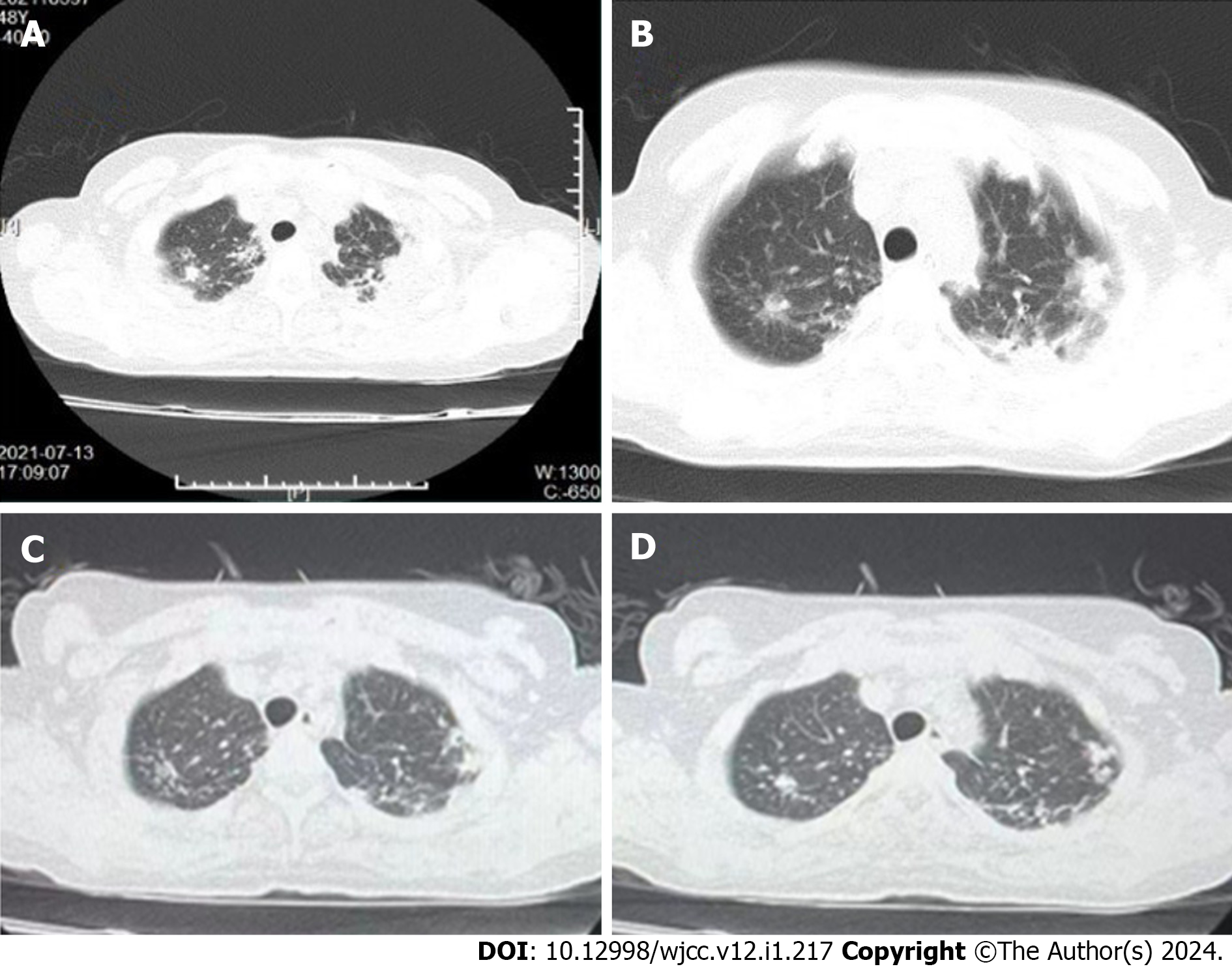
Figure 6 Chest computed tomography scans before and after treatment.
A and B: Before anti tuberculosis treatment; C and D: After 1 mo of treatment, chest computed tomography showed improvement in inflammation.
Figure 7 After 6 mo of treatment, the pigmentation was partially reversed.
A: Hyperpigmentation on the skin of face, mouth, tongue and mucous membranes of the lips; B-D: Hyperpigmentation on nails, hands and feet.
- Citation: Zhang TX, Xu HY, Ma W, Zheng JB. Addison's disease caused by adrenal tuberculosis may lead to misdiagnosis of major depressive disorder: A case report. World J Clin Cases 2024; 12(1): 217-223
- URL: https://www.wjgnet.com/2307-8960/full/v12/i1/217.htm
- DOI: https://dx.doi.org/10.12998/wjcc.v12.i1.217


