
Recommended
More Related Content
What's hot
What's hot (20)
Similar to Sex Cord Stromal Tumors: Granulosa Cell Tumors & Thecoma
Similar to Sex Cord Stromal Tumors: Granulosa Cell Tumors & Thecoma (20)
Recently uploaded
Recently uploaded (20)
Sex Cord Stromal Tumors: Granulosa Cell Tumors & Thecoma
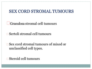
- 1. Granulosa stromal cell tumours Sertoli stromal cell tumours Sex cord stromal tumours of mixed or unclassified cell types. Steroid cell tumours SEX CORD STROMAL TUMOURS
- 2. Granulosa stromal cell tumours (SEX CORD STROMAL TUMOURS) Granulosa cell tumour group a) Adult granulosa cell tumour. b) Juvenile granulosa cell tumour. Thecoma fibroma group a) Thecoma (typical or lutenised) b) Fibroma c) Cellular fibroma d) Fibrosarcoma e) Sclerosing stromal tumours f) Signet ring stromal tumour g) Stromal tumour with minor sex cord elements h) Unclassified
- 3. Granulosa Cell Tumor group (sex cord stromal tumor) Differentiation towards follicular granulosa cells that can occur in adults (adult granulosa cell tumor) and in younger patients (juvenile granulosa cell tumor). Two distinct types: * adult * juvenile
- 4. Adult Granulosa Cell Tumor Usually childbearing age. - 75% have hyperestrinism, which may result in: +isosexual precocious puberty +proliferative breast disease, endometrial hyperplasia and endometrial caecinoma. Elevated serum inhibin and follicle regulatory proteins.
- 5. AGCT The tumors are usually large (>10 cm) and unilateral. The cut surface is soft and yellow-tan with cysts and hemorrhage. Cut surface: -predominantly solid May be: cystic: -filled with straw-colored or mucoid fluid -sometimes so prominent as to simulate appearance of a cystadenoma Granulosa cell tumor with solid cut surface.
- 6. AGCT Different histologic patterns occur, including microfollicular, macrofollicular, nested, cords, gyriform, and diffuse. However, all are composed of round to oval granulosa cells that have little cytoplasm and round to angular nuclei with longitudinal nuclear grooves (coffee bean appearance) There is minimal cytologic atypia. Mitotic rate is low.
- 9. The microfollicular and diffuse variants often contain characteristic Call–Exner bodies consisting of a very small collection of eosinophilic material lined by well-differentiated granulosa cells.
- 10. Cells may be luteinized (plump with ample cytoplasm), particularly during pregnancy; may have theca cell component. Positive stains: Inhibin alpha, vimentin, calretinin, CD99, smooth muscle actin, desmoplakin, S100 (50%), keratin (dot-like in 30-50%). - Silver stains demonstrate reticulin surrounding cluster of cells. Negative stains: EMA
- 11. Inhibin
- 12. Juvenile Granulosa Cell Tumor More aggressive than adult GCT More likely to produce distant metastases ≈80% during first two decades of life * Usually presents with isosexual precocity * Occasionally associated with: - enchondromatosis (Ollier's disease) - Maffucci's syndrome
- 13. Juvenile Granulosa Cell Tumor -Diffuse or macrofollicular patterns of growth - eosinophilic mucin-positive intrafollicular secretion. - larger tumor cells with extensive luteinization - paucity of nuclear grooves - nuclear atypia - variable but often high mitotic activity. The follicle-like spaces seen on low-power examination are a common feature of this neoplasm.
- 14. On high power the tumor cells lack the coffee-bean nuclei seen in the adult type
- 17. Special Stains and Immunohistochemistry granulosa cell tumors Adult Granulosa Cell Tumor * Immunohistochemically: - steroid production: + by both theca and granulosa cells with predominance of: # estradiol in granulosa cell # progesterone in luteinized theca cells - vimentin - desmoplakin (desmosomal plaque protein) - inhibin (also JGCT) follicle regulatory proteins - CD99 keratin: + 33–50% of cases + typical dot-like distribution + mainly CK8 and CK18 types25,26 smooth muscle actin: + nearly all cases S-100- 50% cases Strong immunoreactivity for inhibin in granulosa cell tumor.
- 19. Thecoma Postmenopausal women. Symptoms of hyperestrogenism. Most are unilateral and can measure up to 10 cm in diameter.
- 20. Thecoma Usually unilateral Well-defined capsule Firm consistency Cut surface: White to yellow color * largely or entirely solid * may be cystic Cut surface of thecoma showing a predominance of yellow areas alternating with whitish foci
- 22. THECOMA Fascicles of spindle cells with: centrally placed nuclei moderate amount of pale cytoplasm. Only mild atypia and rare mitoses Intervening tissue may show considerable collagen deposition with focal hyaline plaque formation. Degree of cellularity varies considerably Some thecomas in young women are heavily calcified
- 25. Special Stains and Immunohistochemistry Oil red O: (require fresh tissue) - abundant intracytoplasmic neutral fat Positive for inhibin expression Reticulin stain: Demonstrates reticulin fibers surrounding individual cells (note: reticulin surrounds clusters of cells in granulosa cell tumors) Estradiol usually limited to a small number of tumor cell
- 26. FIBROMA The most common type of sex-cord stromal tumor developing from specialized ovarian stroma. Usually unilateral Almost invariably after puberty Fibromas are not hormonally functional average of 5 cm in diameter Sometimes in young women with basal cell nevus (Gorlin's) syndrome. Benign May be ascites: especially if large sometimes with right-sided pleural effusion (Meigs' syndrome) (disappears on removal of tumor).
- 27. Gross Pathology Cut surface: Solid, firm, white, lobulated Outer aspect of ovarian fibroma
- 29. FIBROMA Spindle stromal cells: - closely packed - arranged in 'feather-stitched' or storiform pattern - no atypia and few mitoses May be: - hyaline bands - edema If in basal cell nevus (Gorlin's) syndrome: - calcified - usually bilateral - often multinodular Cellular fibroma. The tumor is hypercellular, but pleomorphism and mitotic activity are minimal
- 30. Immunohistochemistry -WT1, FOXL2, vimentin, CD56, SF1 positive -SMA, CD34, desmin, ER, PR may be positive Inhibin and calretinin rarely focally positive -CD10 typically negative Genetic Testing -No FOXL2 mutations -Trisomy &/or tetrasomy 12 -Trisomy 8 (fibrosarcoma)