Cryptococcosis
•Download as PPTX, PDF•
69 likes•32,481 views

Cryptococcosis also called as Torulosis is a subacute or chronic fungal infection caused by Cryptococcus neoformans. It leads to compications such as fatal meningoencephalitis. It is an opportunistic infection in HIV-infected patients. The PPT discuss on the morphology of the fungus, pathogenesis, laboratory diagnosis and treatment.

Recommended
More Related Content
What's hot
What's hot (20)
Similar to Cryptococcosis
Similar to Cryptococcosis (20)
More from Mary Mwinga
Recently uploaded
Recently uploaded (20)
Cryptococcosis
- 2. Def: cryptococcoccosis is a subacute or chronic infection caused by yeast Cryptococcus neofomans. Also called Torulosis Produces potentially fatal meningoencephalitis in HIV patients. Causative agents Two species: C. neoformans and C. gattii. Four serotypes: A, B, C and D.
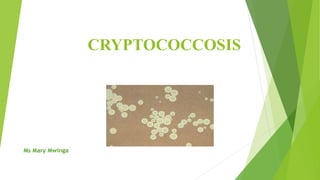
- 3. 1. Cryptococcus neoformans It is a round or ovoid budding cell Size: 4-20 µm in diameter. It’s a true yeast Due to prominent polysaccharide capsule. Varieties of C. neoformans a. C. neoformans var. grubii b. C. neoformans var. neoformans 2. Cryptococcus gattii – is antigenically diverse - corresponds to serotypes A and D.
- 4. Other species: C. albidus and C. laurentii. Other agents of cryptococcosis: Teleomorphs for fungus -belong to Basidiomycetes: Filobasidiella neoformans and F. basiliospora.
- 5. pathogenesis Source: birds’ excretions. Route: Infection is acquired by inhalation of aerosol forms of Cryptococcus through lungs. Leads to pulmonary infection. Other way: through skin or mucosa. Both yeast cells and basidiospores (sexual stage of Cryptococcus) are infectious. In immunocompetent individuals: lungs have defence mechanisms which limit the infection. In people with low immunity: pulmonary infection occurs followed by dissemination through blood.
- 7. CNS spread Cryptococcus has ability to cross the blood-brain barriers. The cells migrate directly across endothelium or carried inside macrophages as ‘Trojan horse’. Present as discrete nodules- Cryptococcoma. Virulence factors Polysaccharide capsule: -is anti-phagocytic. - inhibits hosts local immune responses. Ability to make melanin: - produces an enzyme (phenyl oxidase) - it breaks down caffeic acid to melanin Other enzymes: ex, phospholipase and urease.
- 8. Risk factors Patients with advanced HIV infection. -they have less CD4 T cell counts [<200/µl] -they are at high risk. Patients with haematological malignancies. Transplant recipients. Patients on immunosuppressive or steroid therapy. Old buildings- exposure to spores.
- 9. Clinical manifestations 1. Pulmonary Cryptococcosis: - Respiratory tract: most common entry. - Seen in immunocompetent host. - Patient develops asymptomatic or mildly pneumonitis. - Results in an encapsulated lung nodule: Cryptococcoma. - Symptoms: chronic cough, low grade fever, chest pain, scant mucoid or blood- tinged sputum, malaise (disconfortness) and weight loss
- 10. 2. Disseminated infections May lead to visceral, cutaneous, meningoencephalitis disease or ocular cryptococcosis. A. CNS Cryptococcus/ Cryptococcus meningoencephalitis Present as chronic meningitis C. neoformans var. neoformans and C. gattii are strongly neurotropic -they disseminate from primary pulmonary site to the CNS. - Infection may extend to brain: forms massive lesions or mucoid cysts. - Leads to cryptococcal meningoencephalitis/meningitis. - they invade the leptomeninges. Signs & symptoms: headache, fever, meningismus, loss of vision, sensory & memory loss, and seizures. - Cryptococcal infection mimic tuberculosis and other chronic types of meningitis. - seen in AIDS patients.
- 11. B. Visceral Cryptococcus/osseous cryptococcosis: -simulate tuberculosis and cancer. -leads to osteolytic of bones (osteomyelitis). -uncommon but severe infection. -infection acquired by haematogenous spread from a self-limited pulmonary or lymph node localization, or -originates from contiguous skin lesion.
- 12. C. Cutaneous Cryptococcus -Results from haematogenous dissemination of infection or -Primary cutaneous lesion- following inoculation of the fungus into skin. - Lesions may be papules, acneform pustules or subcutaneous abscesses- may ulcerate. - Ulcers may multiply and resemble carcinoma. - Commonly caused by neoformans species.
- 13. D. Ocular Cryptococcal Patients develop keratitis, papilledema, scotoma, chorioretinitis and ocular palsy Leads to visual loss
- 14. Laboratory diagnosis Specimen collection -specimens: Sputum, CSF, Blood, skin scrapings. 1. Microscopy Negative staining: India Ink and Nigrosin stain -modified India ink with added 2% mercurochrome is used -demonstrates capsule: appears as refractile delineated clear space around the cells. -drawback: India ink is less sensitive (60-70%). KOH Preparation: used for sputum Gram stain: reveals Gram- positive, budding yeast cells. - surrounded by a halo or clear area- reveals capsule.
- 16. Other stains Mucicarmine stain: stains carminophilic cell wall of C. neoformans. Masson- fontana stain: demonstrates production of melanin. Alacin blue stain: demonstrates capsule.
- 17. 2. culture Specimen: inoculated on SDA. Plates are incubated at 37°C. Blood: inoculated in biphasic blood culture bottles. Colonies: mucoid creamy white colonies -cream colour becomes tannish -flat or slightly heaped, shiny, smooth edges
- 18. Other media Inositol agar with chloramphenicol - inhibits Candida growth -used for inoculation of urine and pallets from centrifuged bronchial secretions -inositol is a unique carbon source -assimilated by Cryptococcus spp. -incubation: 3-5 days colonies- do not produce hyphae or pseudo-hyphae.
- 19. Niger seed agar, caffeic acid agar and bird seed agar -demonstrate melanin production: brown coloured colonies - C. neoformans breaks down caffeic acid to melanin -Growth at 37°C. 3. Biochemical confirmation -urease test: positive. -assimilation of inositol, maltose, sucrose, dextrose, galactose, xylose and nitrate. - Not fermentative
- 20. 4. Mouse pathogenicity test -inoculation of colonies in mice: intracerebral or intraperitoneally. -fatal for mice. -Capsulated budding yeast cells: demonstrated in the brain of infected mice.
- 21. 5. Immuno-serology Ag detection - Detects polysaccharide antigen of C. neoformans in fluids. - High titre in serum, intermediate in CSF and lowest in urine. - Bronco- alveolar lavage (BAL): also tested for Cryptococcal Ag. Latex agglutination Test (LAT) - Slide agglutination test: uses latex particles coated with polyclonal or monoclonal antibodies. - Positive test: is done at dilution 1:4 - Titre ≥ 1: 8 indicates active disease. - Higher Ag titre: indicate severe infections - Falling titre: good prognostic sign.
- 22. Treatment Amphotericin B 5- fluorocytosine imidazoles (miconazole, keratonazole) Triazoles (itranazole, fluconazole, voriconazole) Echinocandins (caspofungin, micafungin)
- 23. THANK YOU