
Recommended
More Related Content
What's hot
What's hot (20)
Viewers also liked
Viewers also liked (20)
Similar to Omphalocele vs gastroschisis
Similar to Omphalocele vs gastroschisis (20)
Recently uploaded
Recently uploaded (20)
Omphalocele vs gastroschisis
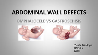
- 1. ABDOMINAL WALL DEFECTS OMPHALOCELE VS GASTROSCHISIS Rusila Tikoitoga MBBS 4 2016
- 2. OBJECTIVES • Background • Epidemiology • Etiology • Pathophysiology • Clinical Features • Diagnosis • Management • Prognosis
- 3. History • 1634 - Ambroise Paré (French barber surgeon) first described Omphalocele. • Derived from Latin word “Omphalos” meaning prominence or navel. • 1733 – James Calder (Scottish neonatal surgeon) first described Gastroschisis. • Derived from the Greek word “Gaster”(Gastro) meaning belly and “schisis” meaning to tear or split from "
- 4. Epidemiology Gastroschisis Incidence - 4 per 10,000 M:F is 1:1 • 10-15% association with congenital anomalies such as CHD(VSD), cleft palate and intestinal atresia • 40% are premature/SGA Omphalocele Incidence - 3 per 5,000 M:F is 1.5:1 >70% association with congenital anomalies such Bowel atresia, Imperforated anus, Trisomies 13, 18, 21, Beckwith-Wiedemann Syndrome & Pentalogy of Cantrell
- 5. Etiology • Gastroschisis o Congenital abdominal wall defect towards the right side of the umbilicus and protruded bowel is not covered by a membrane. o Failure of migration and fusion of the lateral folds of the embryonic disc on the 3rd-4th week of gestation. o Disruption of the right omphalomesenteric artery as midgut returns to abdomen by the 10th week causing ischemia of the abdominal wall and weakness then herniation. o Rupture of omphalocele • Omphalocele o Congenital abdominal wall defect with protrusion of abdominal viscera contained within a parietal peritoneum and amniotic membranous sac with Wharton’s jelly. o Due to failure of the midgut to return to abdomen by the 10th week of gestation during midgut rotation.
- 6. Risk Factors Omphalocele • Increased maternal age • Twins • High gravida • Consecutive children Gastroschisis • Young maternal age • Low gravida • Prematurity • Low birth-weight secondary to IUGR
- 11. Clinical Features OMPHALOCELE • central defect of the abdominal wall beneath the umbilical ring. • Defect may be 2-12 cm (Small-<5cm)(Large>8cm) • Always covered by sac • Sac is made of amnion, Wharton’s jelly and peritoneum • The umbilical cord inserts directly into the sac in an apical or lateral position. • Small contains intestinal loops only. Large may involve liver, spleen and bladder, testes/ovary • >50% have associated anomalies GASTROSCHISIS • Defect to the right of an intact umbilical cord allowing extrusion of abdominal content • Umbilical cord arises from normal place in abdominal wall • Opening <=5 cm • No covering sac (never has a sac ) • Evisceration usually only contains intestinal loops • Bowels often thickened, matted and edematous • 10-15% have associated anomalies • 40% are premature/SGA
- 12. Diagnosis • Alpha-feto-protein-synthesized in fetal liver and excreted by fetal kidneys and crosses placenta by 12 weeks. • Elevated maternal AFP - neural tube defects, abdominal wall defects, duodenal or esophageal atresia • 40% false positive rate • Fetal ultrasound after 14 weeks gestation is the confirmatory test.
- 13. Prenatal Ultrasound • Normal umbilical cord insertion site • Small bowel loops seen in the amniotic cavity • No covering membrane over the loops of bowel • Can include stomach and large bowel • Majority occur to the right of the umbilical cord Gastroschisis
- 14. Prenatal Ultrasound • Umbilical cord insertion is typically midline on the mass • Located centrally • Contents are intestinal loops and maybe liver, spleen and gonads. Omphalocele
- 15. Management Perinatal Management • Maternal Screening Fetal Ultrasound = positive findings Alpha-feto-protein elevated = 90% Omphalocele 10% Gastroschisis • Prenatal counselling
- 16. Pre-operative Management • ABC • Heat Management – Sterile wrap or sterile bowel bag – Radiant warmer • Fluid Management – IV bolus 20 ml/kg LR/NS – D10¼NS 2-3 maintenance rate • Nutrition – TPN (central venous line ) • Abdominal Distention – OG/NG tube – urinary catheter • Infection Control Broad-spectrum antibiotics - Ampicillin and Gentamycin • Closure of the Defect
- 17. Omphalocele • Conservative 1. Large omphalocele (10-12cm) apply topical application - Betadine ointment or silver sulfadiazine to the intact sac. 2. Secondary eschar formation and granulation. 3. Healing lasts for 12 months then repaired as ventral hernia. o Primary Closure Small defects (<4cm) excision of the sac and closure of the fascia and skin over the abdominal contents o Mesh patch Medium defects (6-8cm)
- 18. • Post operative care o NICU o Ventilation o Feeding: – Minimal volume o 48 hrs Antibiotics o Hernia dealt with at 1 yr old
- 19. Gastroschisis • Primary closure o If bowel easily reduced • Staged closure o Silo fashioning: Sac excised Silo sewn to rectus fascia/full thickness
- 20. • Post operative care o NICU o Feeding delayed for weeks o Oral stimulation/sucking reflex o Broad spectrum antibiotics
- 21. Long Term Outcomes Omphalocele • Small - recover well • Large: – Gastro-oesophageal reflux - 43% – Majority improve over time – 20% pulmonary insufficiency – Respiratory Infections – Asthma – Feeding difficulties; • 60% with giant omphalocele • May need gastrostomy for feeding – Failure to thrive Gastroschisis • Generally excellent if no atresia • NEC: – 18.5% of neonates more with formula – Bowel loss - short gut syndrome • Cryptorchidism: – 15-30% – Due either being outside/prematurity – Replacement and orchidopexy by 1 yr • 60% have psychosocial stress if umbilicus sacrificed
- 22. Summary
- 23. References: • Up to Date • Medscape • O.P Ghai E.pediatrics • Rudolph’s pediatrics