Download
1 / 32
340 likes | 600 Views
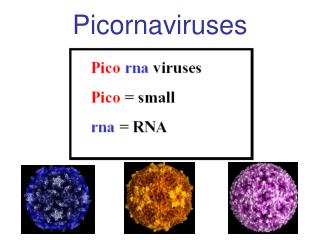
Family: Picornaviridae ( Enteroviruses ). Dr. Mohammed Arif Associate professor Consultant virologist Head of the virology unit. Structure. Picornaviruses = small RNA viruses . Picorna viruses are small, 20-30 nm, icosahedral particles. Unenveloped.

E N D
Family: Picornaviridae ( Enteroviruses ). Dr. Mohammed Arif Associate professor Consultant virologist Head of the virology unit
Structure Picornaviruses = small RNA viruses . Picorna viruses are small, 20-30 nm, icosahedral particles. Unenveloped. The viral genome is ss-RNA, with positive polarity. They replicate in the cytoplasm .
Classification This family is divided into three genera: 1- Genus enterovirus 2- Genus rhinovirus, includes rhinoviruses. 3- Genus hepatovirus, includes hepatitis A virus.
Enteroviruses They are divided into five groups: 1- polioviruses, 3 serotypes. 2- Coxsackie A viruses group A, 23 serotypes. 3- Coxsackie B viruses group B, 6 serotypes. 4- Echoviruses, 31 serotypes. 5- Unclassified enteroviruses, 38 serotypes. More than 100 serotypes .
General characteristics of enteroviruses 1- They are transmitted by the fecal oral route. 2- They are acid stable. 3- They replicate in the pharynx and small intestine. 4- They cause neurological and non-neurological diseases. 5- They shed in stool. 6- Do not cause diarrhea.
Transmission By the fecal oral route: Through contaminated hands. Eating uncooked fruits and vegetables contaminated with infectious fecal material. Drinking water contaminated with infectious fecal material. Contamination of fruits and salads by food handlers.
Endemicity Enteroviruses are endemic in areas with: Low standard of hygiene and sanitation. Primitive sewage system. Absence of proper drinking water-pipe system. Low educational level. Crowded living condition.
Poliomyelitis Caused by polioviruses, three serotypes 1, 2 &3 type 1 is the most paralytogenic. They have no common poliovirus antigen. They share 30-50% homology in the nucleotide sequences. They are enteroviruses. Transmitted by the fecal oral route. Humans are the only natural host for the virus. Poliomyelitis is a disease of infants and young children.
Pathogenecity of poliomyelitis After entry the virus replicates in the oropharyngeal and intestinal mucosa. The virus invades the sub-epithelial tissue and reaches local lymph nodes and blood stream. Primary and secondary viremia occurs. The virus reaches the CNS. Replication occurs in the grey matter particularly the anterior horns of the spinal cord and brain stem. Distinctive ( plaques) produced in the grey matter.
Clinical features IP 7-14 days. Clinically, the disease takes four forms. 1-- Asymptomatic infection: About 95% of infected children develop no symptoms at all. 2-- Minor illness (abortive polio) : about 4-8% of infected children develop fever, nausea, vomiting, malaise, headache and recover completely.
Clinical features 3-- Aseptic meningitis: About 1 % of infected individuals will develop signs and symptoms of aseptic meningitis. Fever, headache, nausea, vomiting and stiffness of neck. Recovery is usual. Paralytic polio: About 0.1 to o.5 % of the infected will suffer from paralytic polio ( flaccid paralysis).
Flaccid paralysis ( paralytic polio). Flaccid paralysis results from viral damage to the motor neurons of the anterior horn of the spinal cord. If damage is severe the paralysis becomes irreversible. Involvement of the medulla may lead to respiratory paralysis and death.
Lab diagnosis. By isolation of the virus in tissue culture, followed by typing the isolated virus. Specimen: feces, rectal swabs, throat swabs.
Prevention 1- live attenuated vaccine( Sabin vaccine) Oral vaccine: Contains the three polioviruses as attenuated strains. They have lost the ability to replicate in the CNS, but can replicate in the gut. They have been attenuated by repeating passage of these viruses in monkey kidney tissue culture.
Prevention The vaccine is administered orally in 3 doses, along with the triple vaccine. Vaccinated children are infectious to others, they shed vaccine strains in feces and saliva, so that vaccine strains circulate in the community.
Advantages of the live attenuated vaccine Induces long lasting immunity. Induces local immunity in the form of IgA production ( gut immunity). Administered orally, without the need of sterile syringes.
Disadvantages of the live attenuated vaccine The only disadvantage of this vaccine is the vaccine strain particular type 3 strain can reverts to virulerence and cause paralysis in those who just been vaccinated. It is estimated that vaccine induced poliomyelitis is seen in rate of 1 in 3000,000 vaccinations.
Prevention 2- Inactivated( killed) vaccine(Salk vaccine): Contains the three polioviruses, which have been inactivated by formaldehyde. The vaccine is given in three injections.
Diseases associated with Coxsackie group A viruses Febrile illness with maculopapular rash. Upper respiratory tract infection. Paralytic disease. Meningitis & encephalitis. Peri and myocarditis. Herpangina. Hand, foot & mouth disease. Acute hemorrhagic conjunctivitis.
Herpangina Caused by group A Coxsackieviruses. Characterized by fever, sore throat, pain on swallowing . Small vesicles appear on the pharynx. Palate uvula and tonsils . Recovery is usual .
Hand, foot and mouth disease . Caused by group A coxsackieviruses . Small vesicles develop on the buccal mucosa, hands and feet . Recovery is usual .
Disease associated with Coxsackie group B viruses. Febrile illness with maculopapular rash. Upper respiratory tract infection. Paralytic disease. Meningitis & encephalitis. Peri & myocarditis. Pleurodynia. Juvenile diabetes/ pancreatitis,
Diseases associated with echoviruses. Febrile illness with maculopapular rash. Upper respiratory tract infection. Paralytic disease. Meningitis & encephalitis. Peri & myocarditis.