

Ovary
Brenner tumors
Benign, borderline and malignant Brenner tumors
Editorial Board Member: Gulisa Turashvili, M.D., Ph.D.
Deputy Editor-in-Chief: Jennifer A. Bennett, M.D.


Last author update: 29 July 2021
Last staff update: 30 January 2024 (update in progress)
Copyright: 2003-2024, PathologyOutlines.com, Inc.
PubMed Search: Ovarian Brenner tumor


Table of Contents
Definition / general | Essential features | ICD coding | Epidemiology | Sites | Pathophysiology | Etiology | Clinical features | Diagnosis | Radiology description | Prognostic factors | Case reports | Treatment | Gross description | Gross images | Frozen section description | Frozen section images | Microscopic (histologic) description | Microscopic (histologic) images | Positive stains | Negative stains | Sample pathology report | Differential diagnosis | Additional references | Board review style question #1 | Board review style answer #1 | Board review style question #2 | Board review style answer #2Cite this page: Huvila J, Gilks CB. Benign, borderline and malignant Brenner tumors. PathologyOutlines.com website. https://www.pathologyoutlines.com/topic/ovarytumorb9brenner.html. Accessed May 6th, 2024.
Definition / general
- Tumor composed of transitional / urothelial-like epithelium, typically embedded in fibromatous stroma
- Benign, borderline and malignant variants are recognized, based on the growth pattern and cytological features of the epithelial cells
Essential features
- Benign Brenner tumor:
- Adenofibromatous architecture with nests of bland transitional epithelium present within fibromatous stroma
- Borderline Brenner tumor:
- Papillary architecture with papillae covered by multilayered transitional epithelium
- There is variable but usually low grade cytological atypia
- Malignant Brenner tumor:
- Stromal invasion by carcinoma with transitional cell features, associated with a benign or borderline Brenner tumor
ICD coding
- ICD-O:
- ICD-11:
- 2F32.Y & XH5DX3 - Brenner tumor, NOS
- 2C73.Y & XH2CH8 - Brenner tumor, borderline malignancy
- 2C73.Y & XH6NJ7 - Brenner tumor, malignant
Epidemiology
- Brenner tumors are most common in the fifth and sixth decades but can occur across a wide age range
Sites
- Ovary
- Rare extraovarian Brenner tumors are reported
Pathophysiology
- Cell of origin of Brenner tumors is controversial; they may arise from Walthard rests
Etiology
- Unknown
Clinical features
- Benign Brenner tumors are usually asymptomatic
- Borderline and malignant Brenner tumors are larger and usually present with findings secondary to an adnexal mass
Diagnosis
- Most benign Brenner tumors are an incidental finding in an ovary removed for other reasons
- Borderline and malignant Brenner tumors are usually diagnosed at the time of removal of an adnexal mass
Radiology description
- Nonspecific findings of a solid or solid and cystic ovarian mass
Prognostic factors
- All reported cases of benign and borderline Brenner tumor have had a benign course, although local recurrence has rarely been reported for the latter (Acta Pathol Microbiol Scand Suppl 1972;233:56)
Case reports
- Benign Brenner tumor:
- 58 year old woman with a coexisting benign Brenner tumor and mucinous cystadenoma (Iran J Pathol 2020;15:334)
- 60 year old postmenopausal woman with ovarian mucinous cystic tumor associated with sarcomatous mural nodule and benign Brenner tumor (Medicine (Baltimore) 2019;98:e14066)
- 68 year old woman with a benign Brenner tumor arising in an ectopic ovary (Int J Gynecol Pathol 2020 Sep 17 [Epub ahead of print])
- Borderline Brenner tumor:
- 68 year old woman with borderline Brenner tumor (J Ovarian Res 2014;7:101)
- Malignant Brenner tumor:
- 62 year old woman with malignant Brenner tumor manifesting as bowel obstruction (BMJ Case Rep 2020;13:e235394)
- 77 year old woman with malignant Brenner tumor (Gynecol Oncol Rep 2017;22:26)
Treatment
- Oophorectomy
- No adjuvant treatment for benign or borderline Brenner tumors
- Adjuvant chemotherapy for advanced stage malignant Brenner tumors
Gross description
- Benign Brenner tumor:
- Small (usually < 2 cm), circumscribed, fibrous tumor with a uniform cut surface
- Calcifications may be present
- Borderline and malignant Brenner tumor:
- Smooth surface, larger (usually > 10 cm) with fleshy, polypoid masses projecting into cystic cavity(s)
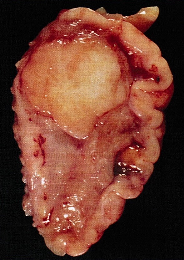
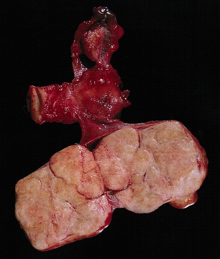
Gross images
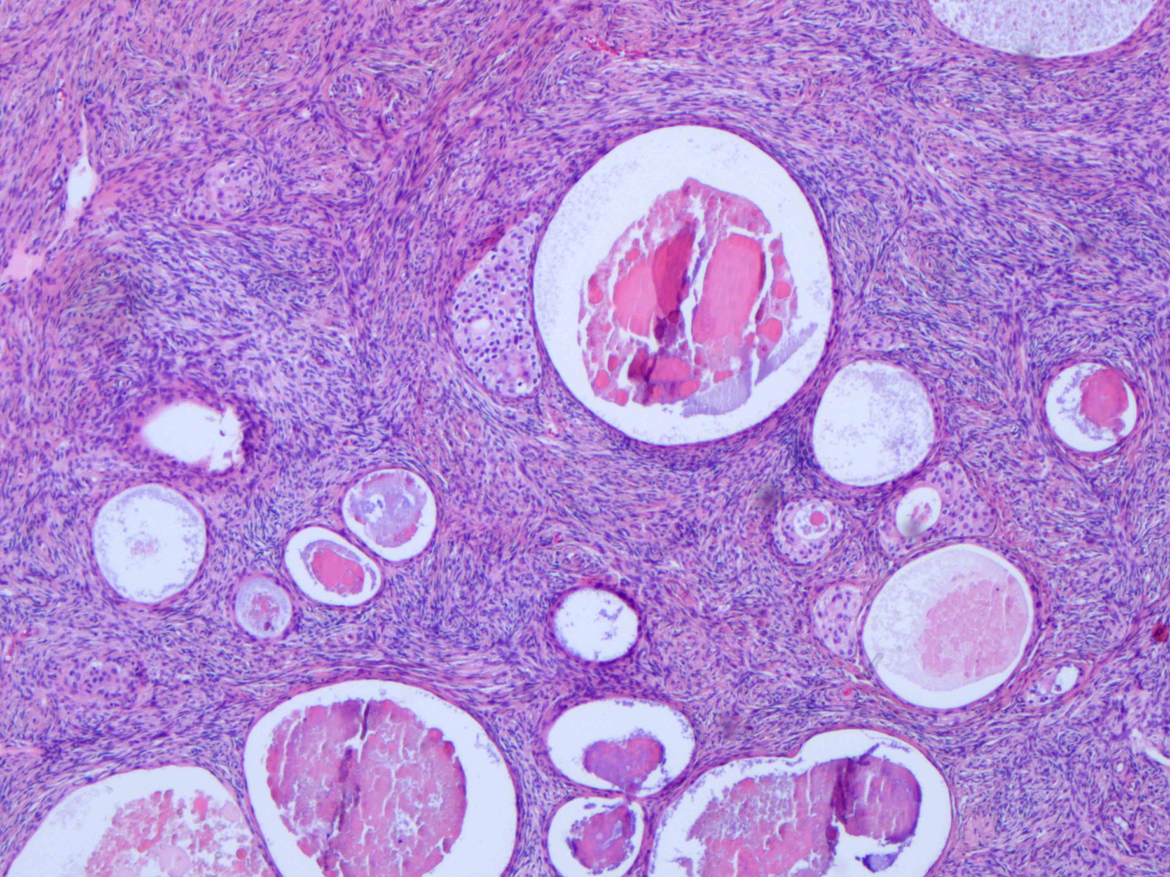
Contributed by Jutta Huvila, M.D., Ph.D. and C. Blake Gilks, M.D. and AFIP images
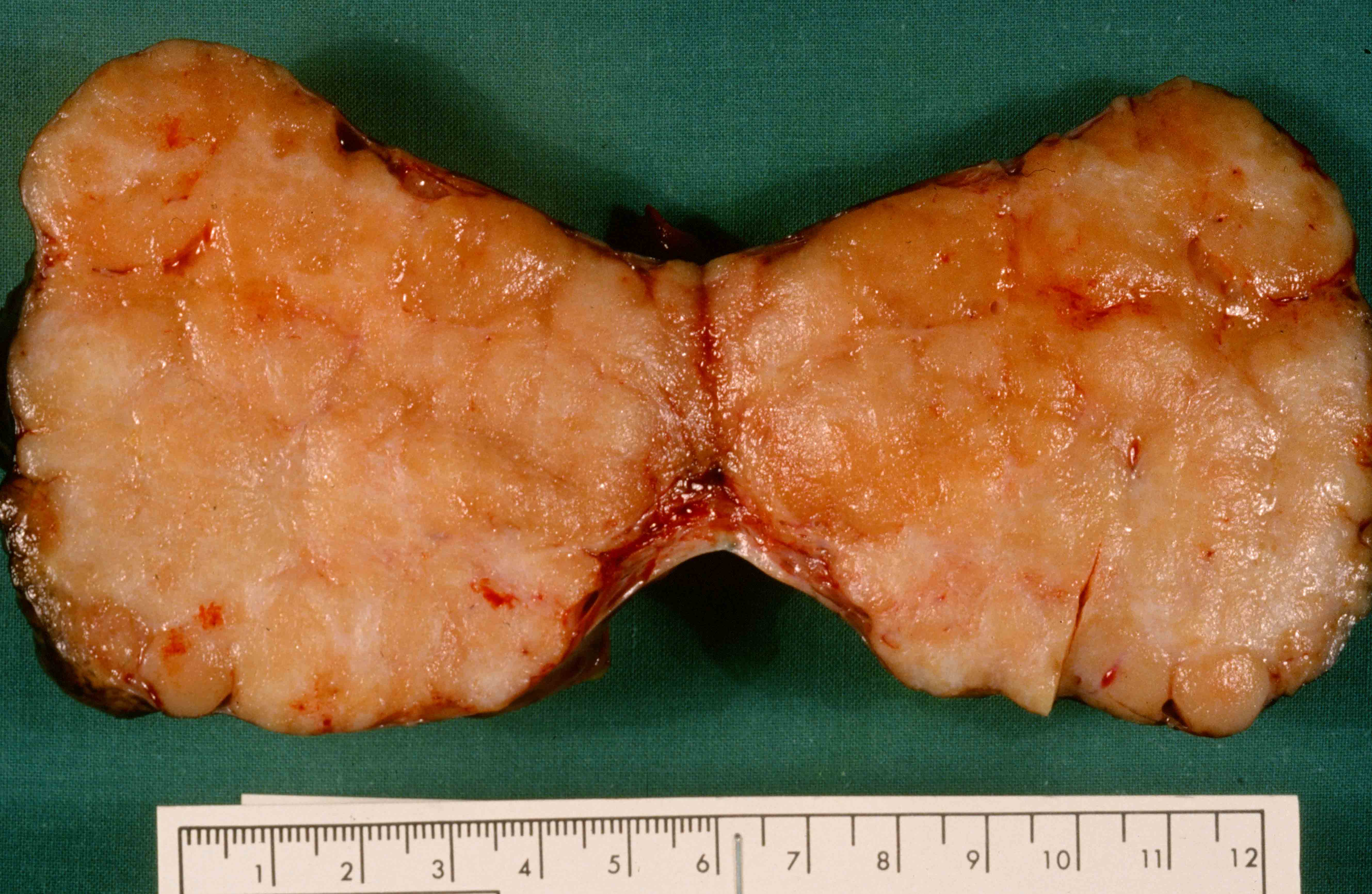
Solid, uniform cut surface

Fleshy appearance
Sharply demarcated
Fibroma-like appearance

Solid fibroma-like component and 2 cysts
Images hosted on other servers:

Infarcted tumor
Frozen section description
- Benign:
- Adenofibromatous architecture, smooth contoured nests of bland epithelial cells within benign fibromatous stroma
- Borderline or malignant:
- Resembling low grade papillary urothelial tumor of the bladder, with papillary fronds covered by transitional-like epithelium
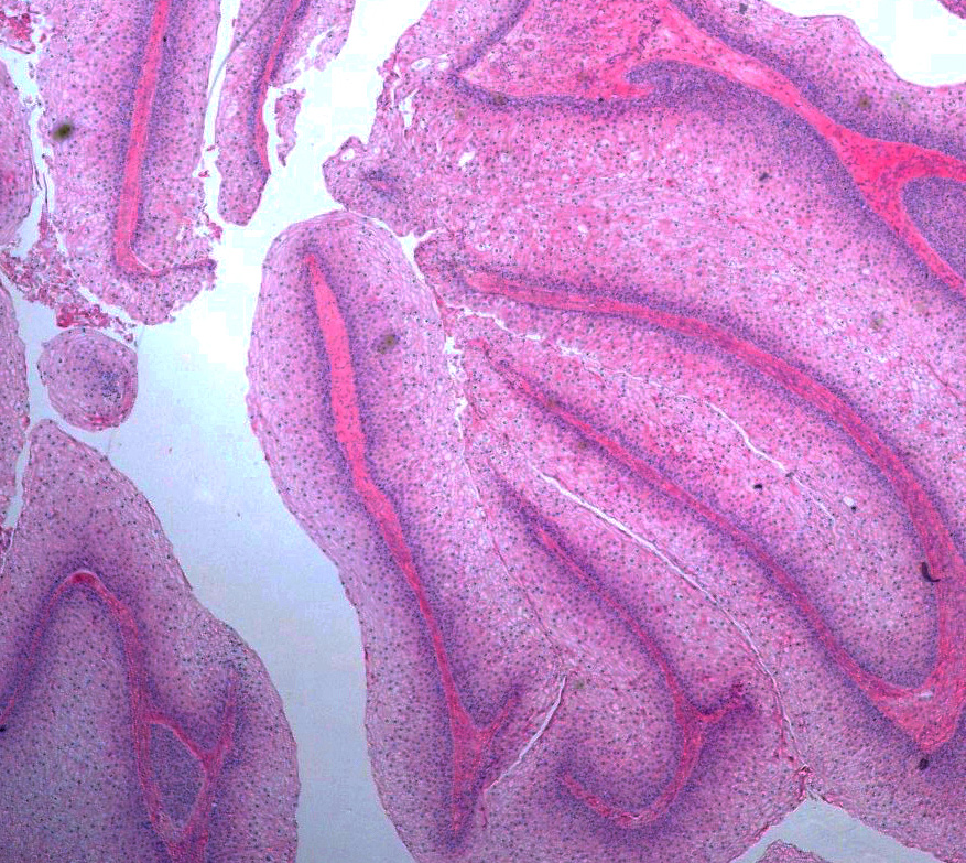
Frozen section images
Contributed by Jutta Huvila, M.D., Ph.D. and C. Blake Gilks, M.D.
Benign Brenner tumor
Microscopic (histologic) description
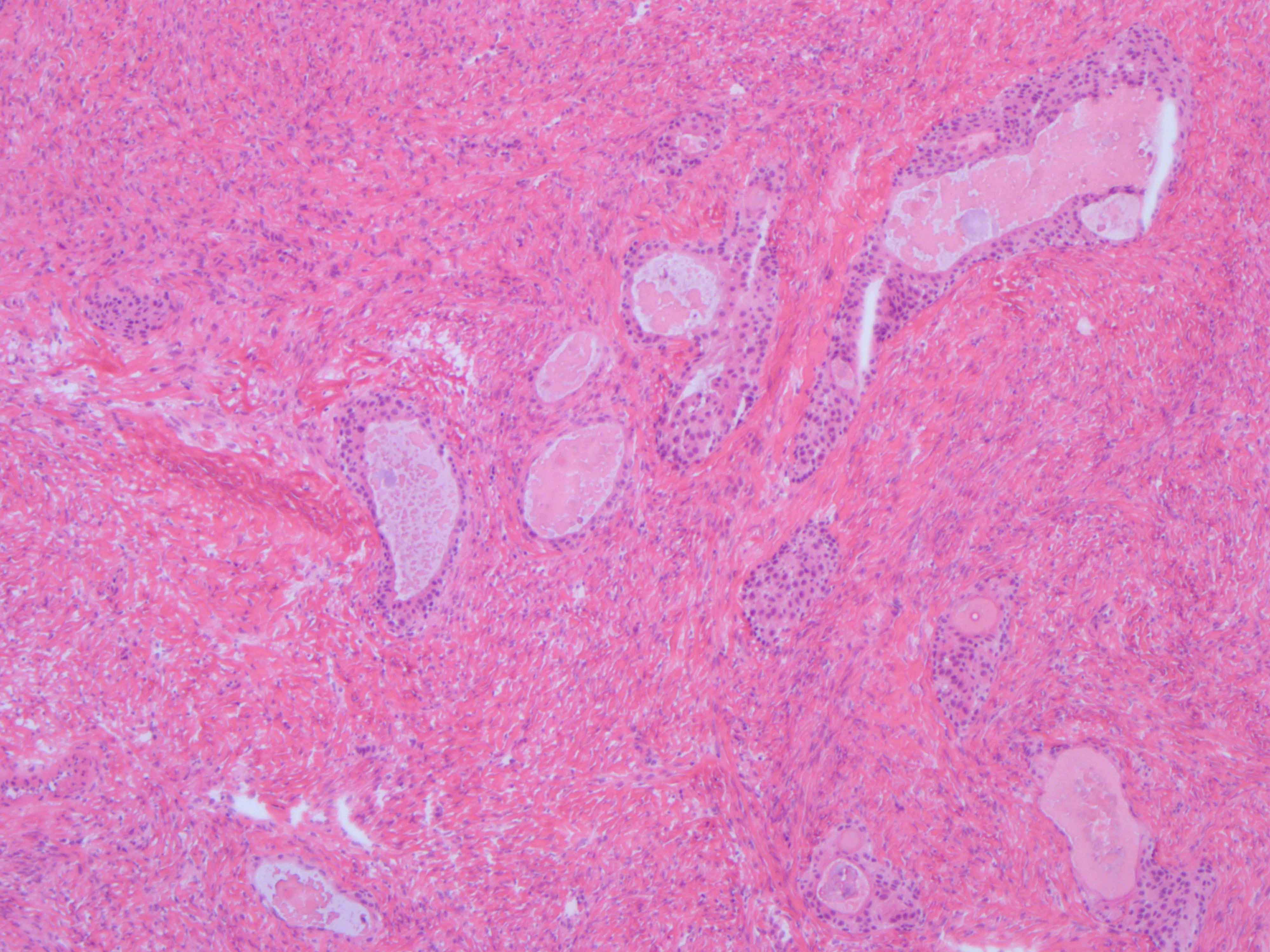
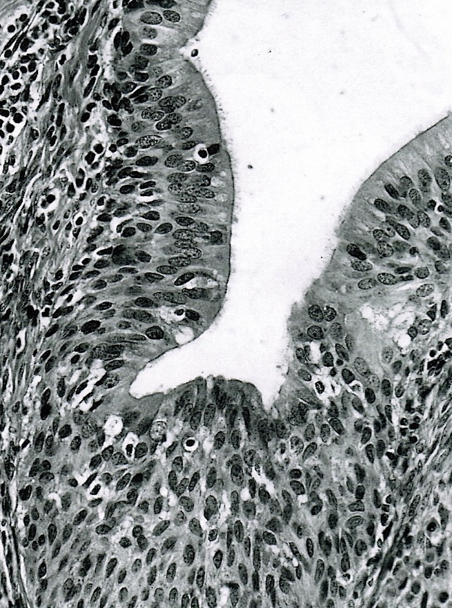
- Benign:
- Smooth contoured nests of bland transitional epithelium within fibromatous stroma
- Transitional cells have uniform oval nuclei and may have a longitudinal nuclear groove
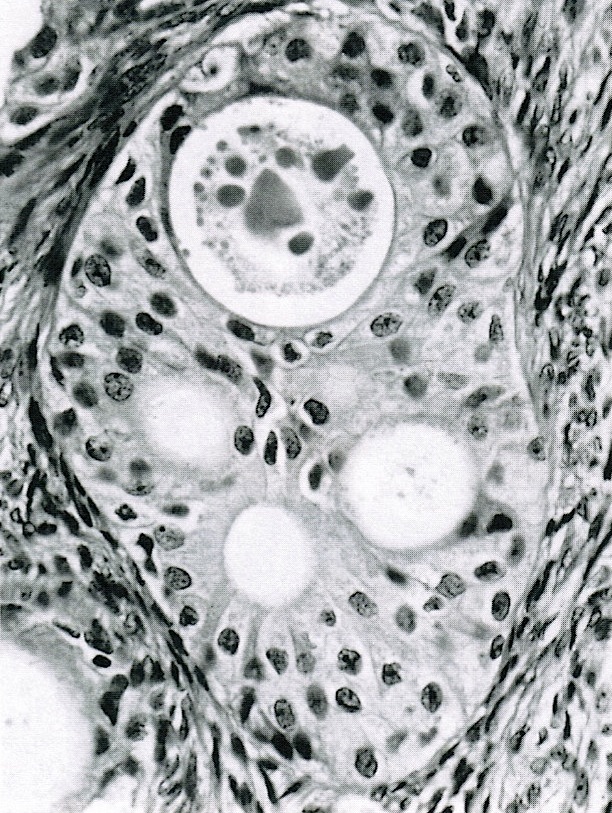
- There may be mucinous epithelium at the center of the nests, with microcyst formation
- Ciliated or nondescript glandular epithelium may be present; a coexistent mucinous cystadenoma is present in 10% of cases
- Calcification is common
- Borderline:
- Papillary architecture with papillae covered by multilayered transitional epithelium
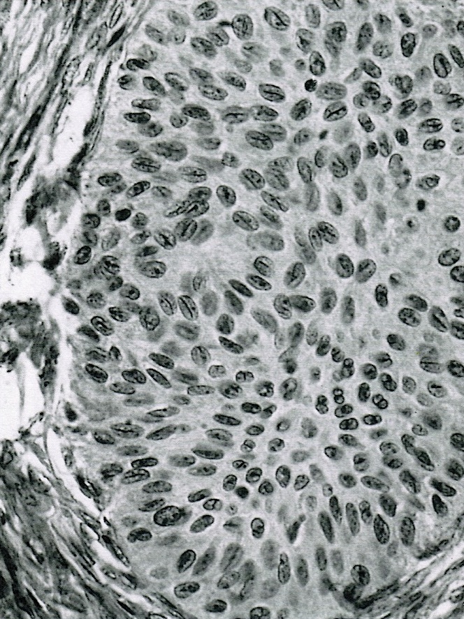
- There is variable cytological atypia; usually low grade but on occasion moderate or marked cytological atypia may be present
- Benign Brenner tumor component is often present
- Malignant:
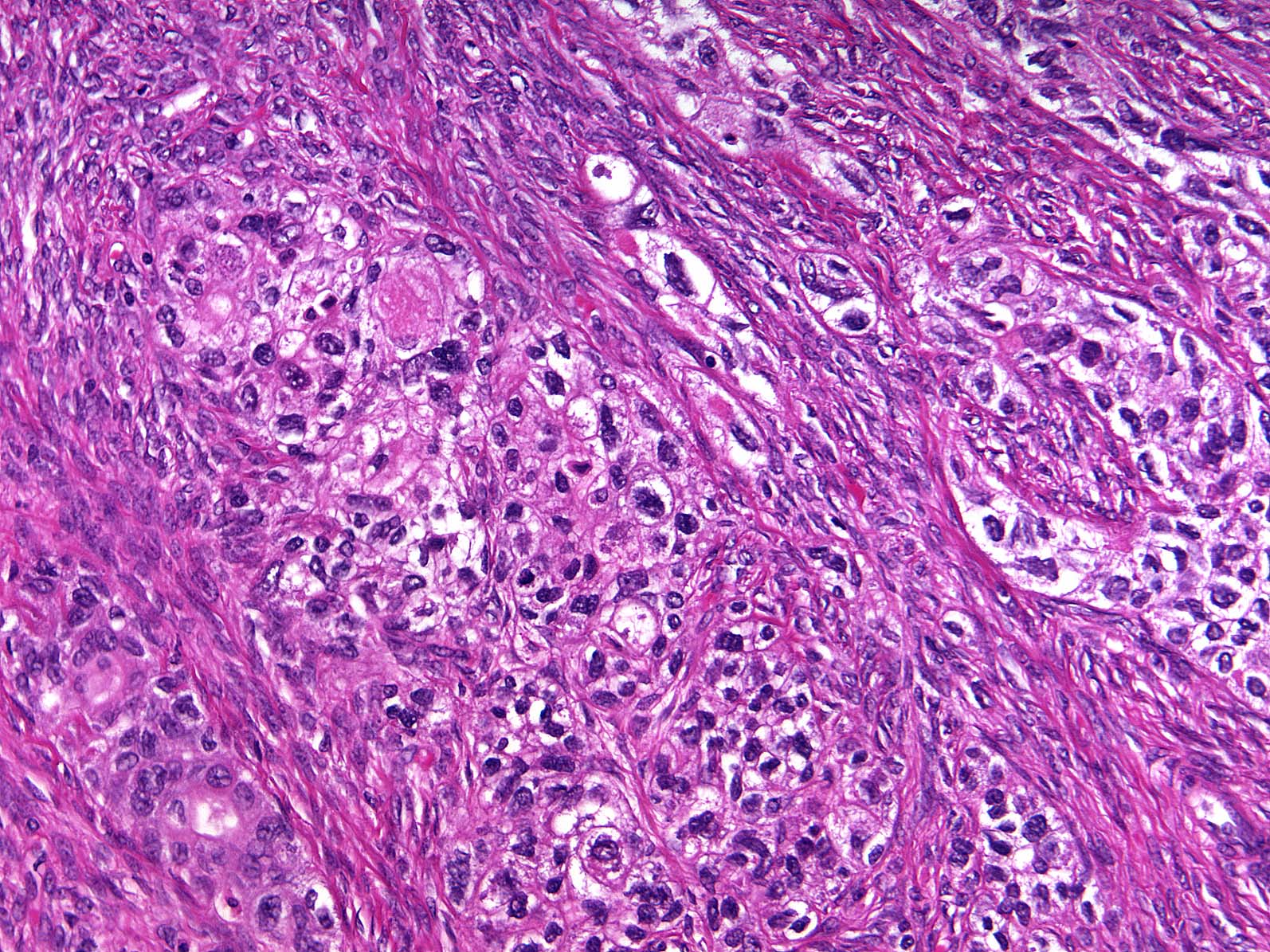
- Stromal invasion by carcinoma with transitional cell features, with irregular nests of cells and single cells in an infiltrative pattern
- Squamous or mucinous differentiation may be present
- Benign or borderline Brenner tumor component is present
- Reference: Int J Gynecol Pathol 2012;31:499
Microscopic (histologic) images
Contributed by Jutta Huvila, M.D., Ph.D. and C. Blake Gilks, M.D.
Benign transitional nests
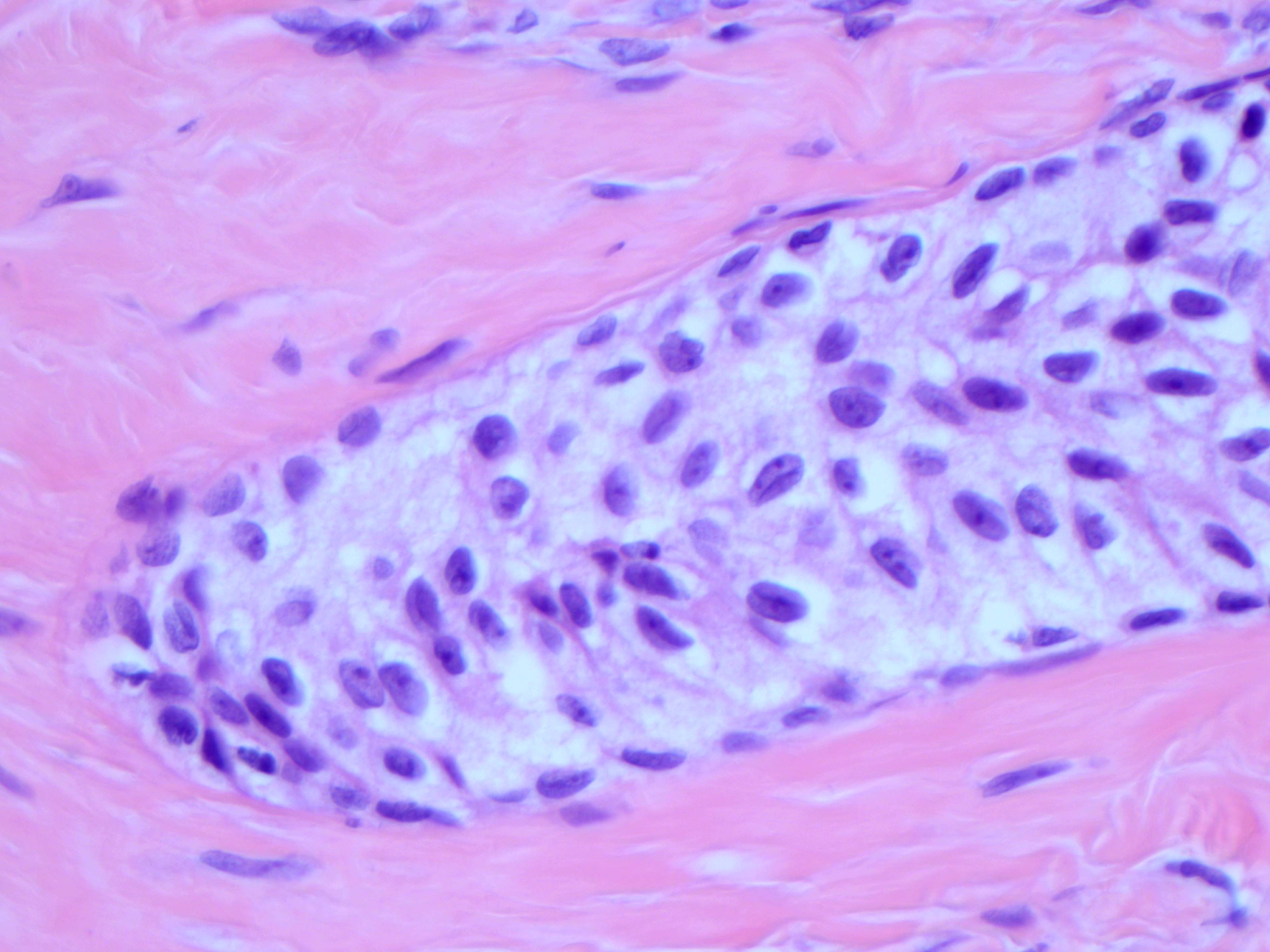
Nuclear grooves
Papillary architecture
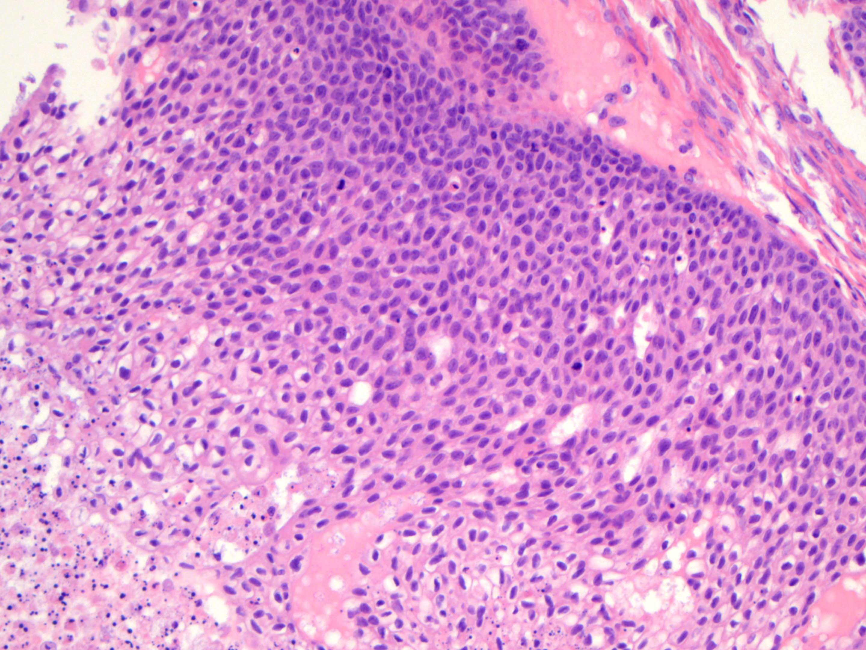
Nuclear atypia
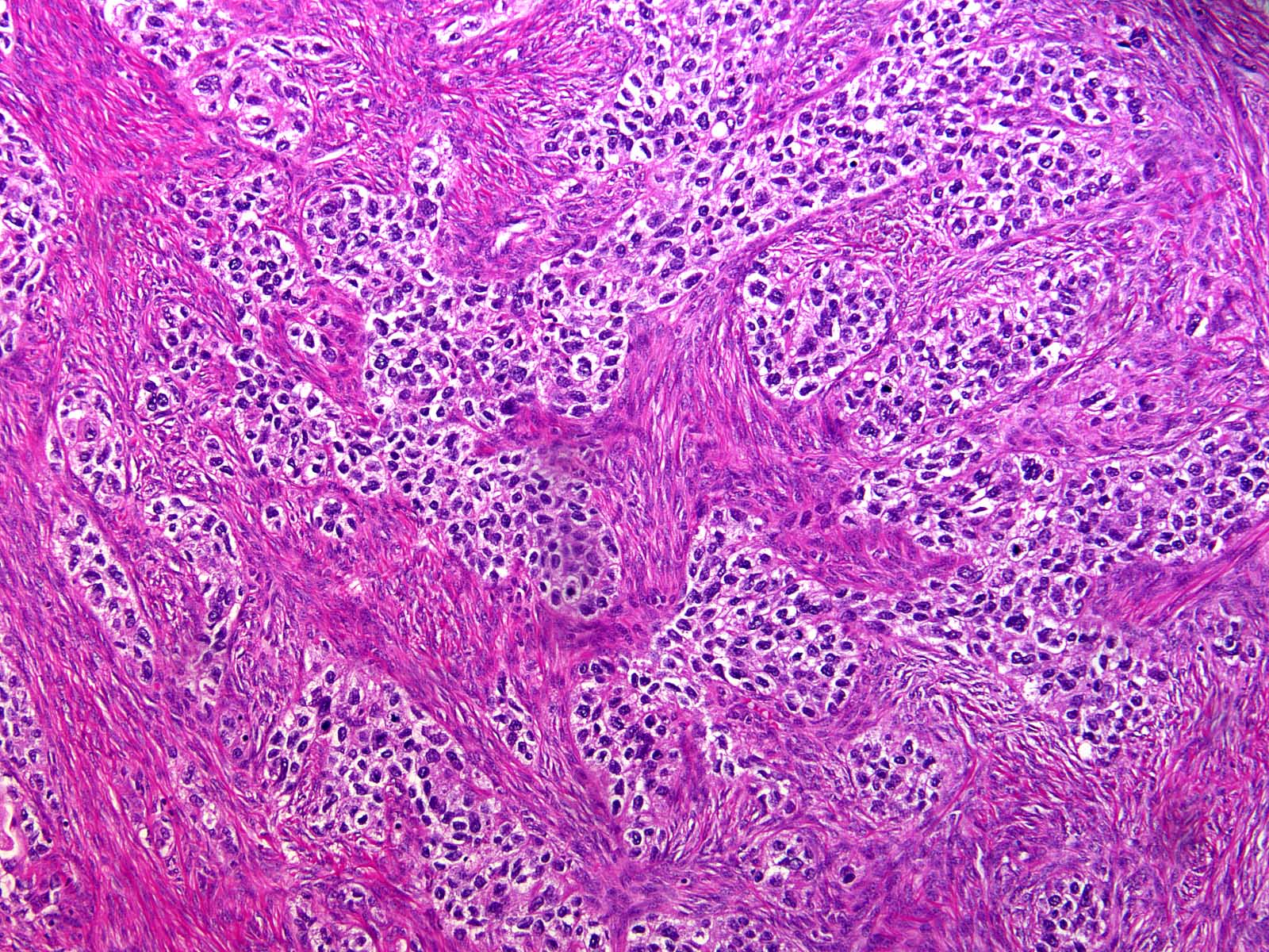
Stromal invasion
Malignant nuclear features
AFIP images

Fibromatous stromal component
Resembling coffee beans
Several lumens
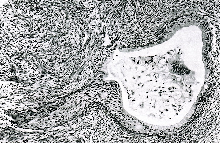
Mucinous epithelium
Ciliated epithelium
Positive stains
Negative stains
Sample pathology report
- Right ovary, oophorectomy:
- Borderline Brenner tumor (see comment)
- Comment: This borderline Brenner tumor is associated with a component of benign Brenner tumor. Negative for invasive carcinoma.
Differential diagnosis
- For Benign Brenner tumor
- Endometrioid adenofibroma:
- Glandular: lacks multilayered epithelial nests with transitional differentiation
- Adult granulosa cell tumor:
- More characteristic patterns of adult granulosa cell tumor present, such as microfollicular, trabecular or solid growth
- Inhibin immunoreactivity and negative for epithelial markers
- Carcinoid tumor:
- Insular or trabecular architecture
- Lacks prominent fibromatous stroma
- Expression of neuroendocrine markers
- Endometrioid adenofibroma:
- For borderline / malignant Brenner tumors
- High grade serous carcinoma, transitional architectural pattern:
- Areas of conventional high grade serous carcinoma
- High grade nuclear features
- Absence of a benign Brenner component
- WT1 and ER positivity (Int J Gynecol Pathol 2012;31:49)
- Squamous cell carcinoma:
- Keratinization
- High grade cytological features
- Teratoma component
- Endometrioid borderline tumor or carcinoma:
- More prominent glandular component with endometrioid (not mucinous) glands
- ER positive
- Metastatic squamous cell carcinoma:
- History of primary squamous cell carcinoma elsewhere in the body
- Bilateral and multinodular growth
- Lacks prominent papillary architecture
- Metastatic urothelial carcinoma:
- History of urothelial carcinoma
- Bilateral and multinodular growth
- Lacks benign Brenner component
- High grade serous carcinoma, transitional architectural pattern:
Additional references
Board review style question #1
Which of the following is true about benign Brenner tumors of the ovary?
- Composed of both epithelium and fibromatous stroma
- Papillary architecture
- Typically > 10 cm
- Usually present with abdominal pain
Board review style answer #1
Board review style question #2
Brenner tumors of the ovary typically have which of the following immunophenotypes?
- GATA3 and ER positive
- GATA3 and p63 positive
- GATA3 and WT1 positive
- GATA3 positive and mutant pattern p53 expression
Board review style answer #2
