by Jason Wasserman MD PhD FRCPC
December 13, 2023
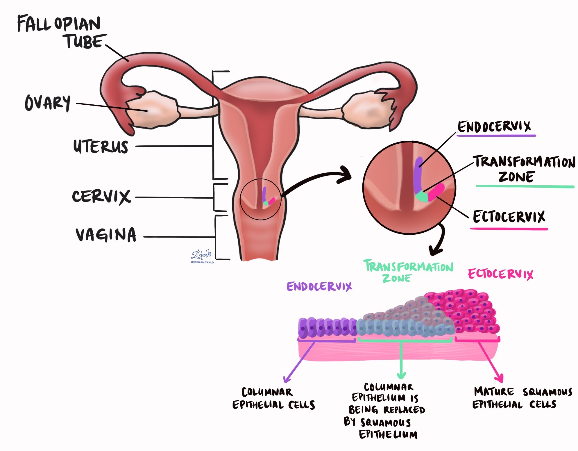
High grade squamous intraepithelial lesion (HSIL) of the cervix is a precancerous disease caused by human papillomavirus (HPV). It is composed of abnormal squamous cells that have been infected and transformed by the virus. The squamous cells are found in a part of the cervix called the transformation zone. There are many types of HPV but most cases of HSIL are caused by the high-risk types 6, 11, 16, 18, and 51. Another name for HSIL of the cervix is cervical intraepithelial neoplasia (CIN).
HSIL is not cancer although patients with HSIL are at increased risk for developing a type of cervical cancer called squamous cell carcinoma. For this reason, most patients with HSIL are offered treatment to remove the area of abnormal tissue. Low grade squamous intraepithelial lesion (LSIL) is a related condition that is also caused by HPV. However, compared to HSIL, the risk of developing cancer from LSIL is much lower.
Your pathology report for high grade squamous intraepithelial lesion of the cervix
The information found in your pathology report for HSIL of the cervix will depend on the procedure performed. For small procedures such as a Pap test, your report may only include the diagnosis. A biopsy report may also include the results of a test called immunohistochemistry performed to look for p16 in the abnormal squamous cells. For larger procedures such as an excision performed to remove the entire area of HSIL, additional information such as the size of the area involved by HSIL and the assessment of margins may also be described. Please see the sections below for more details.
What does high grade intraepithelial lesion look like under the microscope?
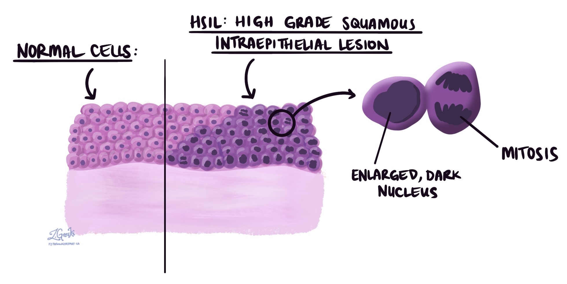
Under microscope examination, the abnormal squamous cells are confined to the epithelium on the surface of the tissue. The abnormal squamous cells are typically larger and darker than the surrounding normal squamous cells. Cells undergoing a process called mitosis (dividing to create new cells) are also usually seen. Pathologists often confirm the diagnosis by performing a test called immunohistochemistry (IHC) for a protein called p16. The abnormal cells in HSIL will be positive for p16 whereas other conditions that can look like HSIL will be negative.
What happens after high grade squamous intraepithelial lesion is diagnosed on a Pap test?
After being diagnosed with HSIL your doctor should refer you to a specialist who will perform a colposcopy. A colposcopy allows your doctor to see the entire outer surface of the cervix. During the colposcopy, the doctor will be looking for any areas that look abnormal on the surface of the cervix. If an abnormality is found, the doctor may decide to take a small biopsy, to confirm the diagnosis of HSIL and to look for squamous cell carcinoma. Your doctor may also take a small sample of tissue from the endocervical canal and endometrium. All patients with HSIL should be followed closely or offered treatment to remove the disease. Talk to your doctor about the options available.
What is a margin and why are margins important?
A margin is any tissue that has to be cut by the surgeon to remove the area of HSIL from your cervix. If you underwent a surgical procedure such as a LEEP, your pathologist will examine the margin closely to ensure there are no abnormal cells at the cut edge of the tissue. Pap smears and small biopsies do not have margins.
A margin is considered positive when HSIL is seen at the edge of the cut tissue. Finding HSIL at the margin increases the risk that the disease will grow back in that location. The number and type of margins described in your report will depend on the type of procedure performed to remove the abnormal tissue from your body.
Typical cervical margins include:
- Endocervical margin – This is where the cervix meets the inside of the uterus.
- Ectocervical margin – This is the bottom of the cervix, closest to the vagina.
- Stromal margin – This is the tissue inside the wall of the cervix.

About this article
This article was written by doctors to help you read and understand your pathology report. Contact us if you have questions about this article or your pathology report. For a complete introduction to your pathology report, read this article.
Related articles on MyPathologyReport
Low grade squamous intraepithelial lesion of the cervix
Squamous cell carcinoma of the cervix
Human papillomavirus (HPV)
Other helpful resources
American College of Obstetricians and Gynecologists
Atlas of pathology

Do you have a question about your pathology report?
Our team of experts is here to help.
Ask a Pathologist.
Our work is generously supported by:
![]()