Case Report

Identifying the Carcinogenic Mechanism of Malignant Struma Ovarii Using Whole-Exome Sequencing and DNA Methylation Analysis

Department of Obstetrics and Gynecology, Shimane University School of Medicine, Izumo 693-8501, Japan
*
Author to whom correspondence should be addressed.
Curr. Issues Mol. Biol. 2023, 45(3), 1843-1851; https://doi.org/10.3390/cimb45030118
Submission received: 30 January 2023 / Revised: 17 February 2023 / Accepted: 21 February 2023 / Published: 23 February 2023
(This article belongs to the Special Issue Next-Generation Sequencing (NGS) Technique and Personalized Medicine)

Abstract

:
Background: Since malignant struma ovarii is a very rare disease, its carcinogenic mechanism has not been elucidated. Here, we sought to identify the genetic lesions that may have led to the carcinogenesis of a rare case of malignant struma ovarii (follicular carcinoma) with peritoneal dissemination. Methods: DNA was extracted from the paraffin-embedded sections of normal uterine tissues and malignant struma ovarii for genetic analysis. Whole-exome sequencing and DNA methylation analysis were then performed. Results: Germline variants of RECQL4, CNTNAP2, and PRDM2, which are tumor-suppressor genes, were detected by whole-exome sequencing. Somatic uniparental disomy (UPD) was also observed in these three genes. Additionally, the methylation of FRMD6-AS2, SESN3, CYTL1, MIR4429, HIF3A, and ATP1B2, which are associated with tumor growth suppression, was detected by DNA methylation analysis. Conclusions: Somatic UPD and DNA methylation in tumor suppressor genes may be associated with the pathogenesis of malignant struma ovarii. To our knowledge, this is the first report of whole-exome sequencing and DNA methylation analysis in malignant struma ovarii. Genetic and DNA methylation analysis may help elucidate the mechanism of carcinogenesis in rare diseases and guide treatment decisions.

1. Introduction

Struma ovarii is defined as a mature cystic teratoma that contains more than 50% thyroid tissue. Struma ovarii accounts for approximately 5% of mature cystic teratomas [1]. The malignant transformation of struma ovarii occurs in only 5–10% of struma ovarii cases. Additionally, the frequency of distant metastases is very low, occurring in only 5–6% of cases [2,3]. Given its rarity, malignant struma ovarii’s carcinogenic mechanism has not been elucidated and no standard treatment has been established. Surgery and 131I therapy have been reported as the main treatment for malignant struma ovarii [4]. However, there have been few reports on the treatment of residual tumors after surgery and 131I therapy. Here, we report a case of malignant struma ovarii with peritoneal dissemination. The patient was treated with surgery, 131I therapy, and chemotherapy. However, the peritoneal dissemination gradually increased. Therefore, genetic analysis was performed to elucidate the cause of malignant struma ovarii and provide the guidance for treatment.

2. Materials and Methods
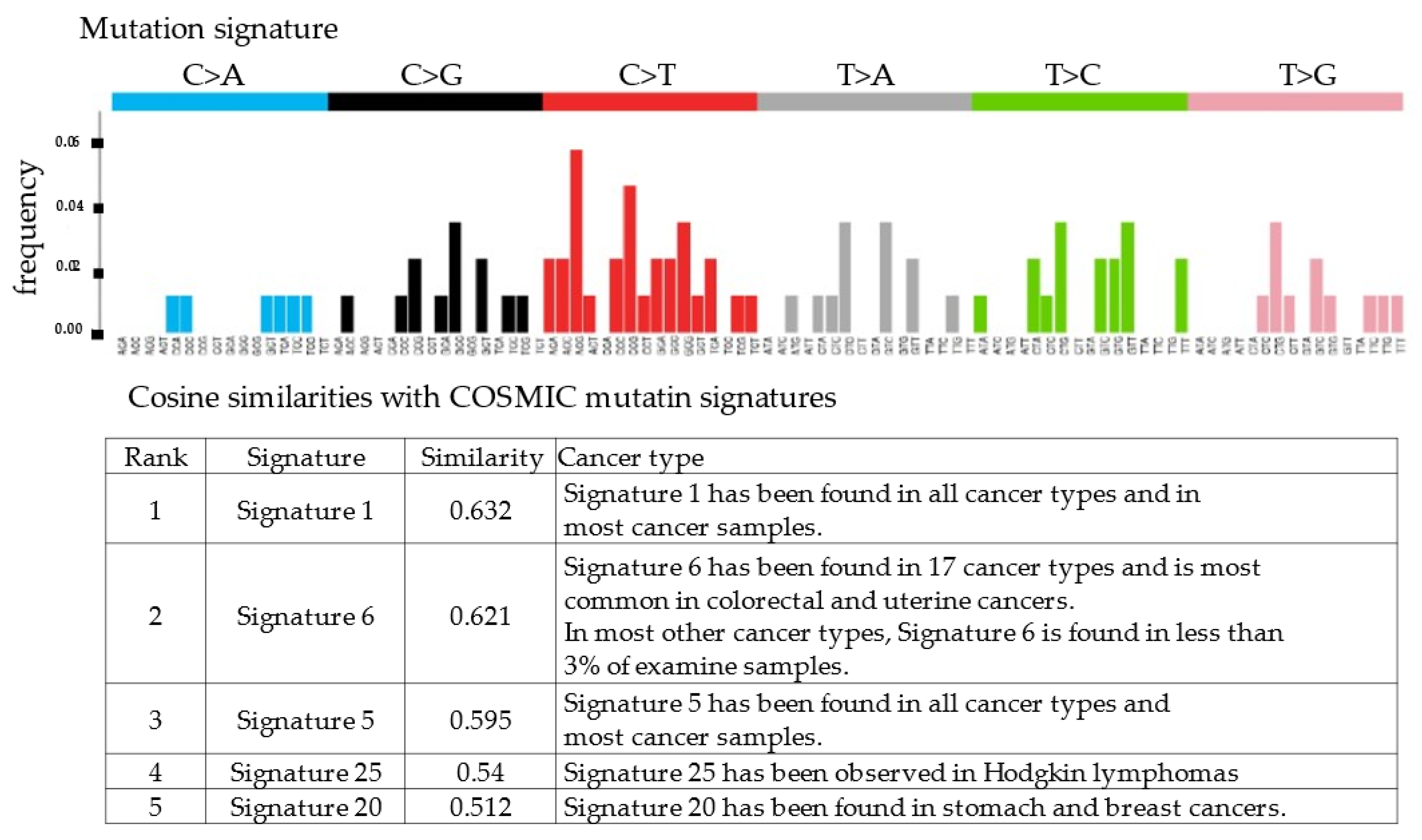
This study was conducted according to the ethical standards of national and international guidelines as well as the Declaration of Helsinki and was approved by the institutional review board (Shimane University Hospital). Tumor specimens were collected after obtaining written consent from the patient with the approval of the Facility Ethical Committee (Shimane University Hospital; approval no. 2004-0381).
Genetic analysis was performed using the PleSSision exome® test (MITSUBISHI SPACE SOFTWARE, Tokyo, Japan) [5]. Hypermutated and ultra-hypermutated statuses were defined as ≥10 and ≥100 SNVs/Mbp, respectively, as previously described [6]. For genetic analysis, DNA was extracted from the paraffin-embedded sections of normal uterine tissues and malignant struma ovarii. The annotation and curation of the sequencing data were performed using the bioinformatics pipeline GenomeJack (MITSUBISHI SPACE SOFTWARE).
We used the Infinium MethylationEPIC BeadChip® (Illumine, San Diego, CA, USA) to analyze the methylation rates of normal uterine tissues and malignant struma ovarii. DNA methylation levels were expressed as β values in normal tissues and malignant struma ovarii. The β value ranges from 0 to 1, and approaches 1 as the methylation rate increases. The difference in methylation rate between normal tissues and malignant struma ovarii for the genes was obtained by the log ratio. The higher the methylation rate in malignant struma ovarii compared to that in the normal tissue, the higher the log ratio. We extracted the top 50 genes with large differences in methylation rates and picked up genes that may have been involved in tumor growth due to methylation changes in malignant struma ovarii.

3. Results

A 29-year-old unmarried, nulliparous woman visited a gynecological clinic due to irregular vaginal bleeding. The patient was referred to our hospital for further examination. She had undergone a laparoscopic left ovarian cystectomy 4 years prior to the visit to our hospital because of a mature cystic teratoma including thyroid tissue.
A multiple cystic mass was detected in the left ovary by transvaginal ultrasonography. To further investigate the left ovarian tumor, magnetic resonance imaging (MRI) and computed tomography (CT) were performed. MRI showed a solid component in the left ovarian tumor, and CT showed peritoneal dissemination (Figure 1A,B). Ovarian cancer was suspected based on the medical examination and imaging findings. Therefore, open surgery was selected as the treatment.
Intraoperatively, the left ovarian tumor was approximately 7 cm in size and adhered to the rectum. Extensive peritoneal dissemination (max. 3 cm) was observed (Figure 2A,B). First, we removed the left ovary. The intraoperative pathological diagnosis of the tumor in the left ovary was malignant struma ovarii (follicular carcinoma). Since the patient elected to not preserve fertility, total hysterectomy, bilateral salpingo-oophorectomy, and omentectomy were performed. After the operation, >2 cm of peritoneal dissemination remained (suboptimal surgery).
The pathological analysis of the resected tumor indicated that the histological type was a highly differentiated follicular carcinoma (Figure 3). No findings of papillary carcinoma, such as psammoma body, intranuclear cytoplasmic inclusion, ground glass nuclei, or nuclear groove, were observed. Similar histology was observed on the posterior wall of the uterus, omentum, and mesentery. This case was diagnosed as Stage IIIC by the 2014 International Federation of Gynecology and Obstetrics (FIGO).
Approximately 8% of patients with malignant struma ovarii are reported to have hyperthyroidism [7], but the patient was euthyroid (thyroid-stimulating hormone, 0.7 µU/mL; free T3, 2.6 pg/mL; free T4, 1.4 ng/dL). Because the ovarian metastasis of thyroid cancer must be ruled out for the diagnosis of malignant struma ovarii, we examined the removed thyroid gland but found no malignant findings. After thyroidectomy, 131I therapy (100 mCi) was used to treat the residual tumor. After 131I therapy, the peritoneal dissemination gradually increased. Therefore, chemotherapy (paclitaxel, carboplatin, and bevacizumab) was administered based on the treatment for the residual ovary tumor. However, the peritoneal dissemination increased slowly during chemotherapy.
Most cases of malignant struma ovarii are only treated with surgery and 131I therapy, and few case reports have shown other types of treatment. Therefore, we performed the genetic and DNA methylation analyses to the identify genetic lesions that may have led to the carcinogenesis of malignant struma ovarii that was intractable even after surgery and 131I therapy.
RecQ like heliccase4 (RECQL4), CNTNAP2, and PRDM2 variants, which are tumor suppressor genes, were detected by whole-exome sequencing. All three variants were germline variants and revealed a somatic uniparental disomy (somatic UPD). Variant positions and variant allele frequencies are shown in Table 1. However, no actionable variants and no copy number alterations were identified in the known oncogenes. The copy number plot was normal and therefore, the tumor was considered homologous recombination proficient (HRP) (Figure 4). Moreover, it was determined to be microsatellite-stable given that the microsatellite instability was 3.82%. The tumor mutation rate was 1.73 SNVs/Mbp, indicating no tumor mutation burden. We also examined the mutation signatures of the malignant struma ovarii. The most prevalent mutational signature was signature 1, which has been found in all cancer types and in most cancer samples (Figure 5).
The DNA methylation analysis detected the methylation of FRMD6-AS2, SESN3, CYTL1, MIR4429, HIF3A, and ATP1B2 in malignant struma ovarii (Table 2). All these genes were reportedly associated with tumor growth suppression; thus, it is possible that the methylation of these genes was involved in the carcinogenesis of malignant struma ovarii [8,9,10,11,12,13,14].

4. Discussion

Malignant struma ovarii is a very rare disease, with only approximately 200 case reports in the published literature. Only 8% of patients with malignant struma ovarii have hyperthyroidism; thus, malignant struma ovarii is rarely diagnosed based on the symptoms of hyperthyroidism [7]. In addition, preoperative diagnosis is very difficult because the MRI findings of malignant struma ovarii are nonspecific. Therefore, malignant struma ovarii is usually diagnosed after surgery. Since malignant struma ovarii is a rare disease, its carcinogenic mechanism is unknown, and no standard treatment has been established. In case reports, surgery, thyroidectomy, and 131I therapy are the most common treatments [4]. Here, we report a rare case of malignant struma ovarii with peritoneal dissemination. The patient was treated with surgery, 131I therapy, and chemotherapy. However, peritoneal dissemination gradually increased. Therefore, we sought to identify the genetic lesions that may have led to the development of malignant struma ovarii, which can provide guidance for treatment. To that aim, we performed genetic and DNA methylation analyses. We detected the germline variants of RECQL4, CNTNAP2, and PRDM2, which are tumor suppressor genes. Furthermore, somatic UPD was observed in these three genes. In addition, the methylation of FRMD6-AS2, SESN3, CYTL1, MIR4429, HIF3A, and ATP1B2, which are associated with tumor growth suppression, was also detected. These findings suggest that somatic UPD and DNA methylation in tumor suppressor genes may be associated with the carcinogenesis of malignant struma ovarii.
Papillary carcinoma is the most common histological subtype of malignant struma ovarii, and follicular carcinoma, as seen in the present case, is rare [15]. Total thyroidectomy is recommended to confirm the diagnosis of malignant struma ovarii by excluding ovarian metastases from primary thyroid cancer to improve the effectiveness of 131I therapy by allowing preferential uptake into residual struma or metastasis [16].
Previous reports have described different surgical procedures for malignant struma ovarii. In a population-level analysis of 68 patients, 33.0% of patients underwent unilateral oophorectomy, 28.6% underwent bilateral oophorectomy, 28.6% underwent oophorectomy and omentectomy, and 4.8% underwent debulking surgery [4]. The average age of the 68 patients was 43 years, and the overall survival rates at 5 and 10 years were 96.7% and 94.3%, respectively [4]. The 5-year and 10-year recurrent rates of the patients with MSO confined to the ovary were 27.1% and 35.2%, respectively [17]. Although the survival rate of malignant struma ovarii is relatively good, it is unclear whether conservative surgery is possible in young patients with peritoneal dissemination, as in the present case. The large tumor size (≥10 cm), adhesion to the surrounding organs, ascites (≥1 L), capsule rupture, stromal component higher than 80%, extensive papillary carcinoma, especially with solid components, necrosis, and more than 5 mitoses per 10 high-power fields have been reported as factors that predict malignancy [18]. Follicular cancer may be more susceptible to hematogenous metastasis than papillary cancer, as it is observed in primary thyroid carcinoma [19,20]. If more cases of malignant struma ovarii are accumulated, the prognosis can be predicted better, and treatment guidelines may be determined based on the prognosis prediction. Conservative surgery may be acceptable in cases without poor prognostic factors.
Very few studies have reported the treatment of malignant struma ovarii by means other than surgery and 131I therapy. However, one case report described a patient with lung and bone metastases who survived more than 20 years after treatment with tegafur-uracil, paclitaxel/carboplatin, and oral etoposide [19]. In malignant tumors for which standard treatment has not been established or that are resistant to conventional treatments, genetic analysis using next-generation sequencing is now being used to guide treatment decision making. Associations between histological types and genetic variants were reported in primary thyroid carcinoma. BRAFV600E, RAS, and rearranged during transfection/papillary thyroid carcinoma (RET/PTC) variants are common in primary papillary thyroid carcinomas [21,22]. In primary PTCs, RET/PTC rearrangements correlate with young age, tumor morphology, and a high frequency of lymph node metastases [23]. BRAF variants have been reported to be associated with aging, advanced disease, classical papillary histology, and poor prognosis [24,25]. Conversely, in primary follicular thyroid carcinomas, RAS variants and PAX8/Peroxisome Proliferator-Activated receptor γ (PPARγ) fusions are found in approximately 80% of cases [26].
Several genetic variants have been reported in malignant struma ovarii. RET/PTC1 and RET/PTC3 rearrangements have been observed in follicular variant PTC originating from struma ovarii [27]. BRAFV600E, KRAS variants, NRAS variants, and HRAS variants have also been observed in papillary carcinoma originating from struma ovarii [28,29,30,31,32,33]. Primary PTC and malignant struma ovarii with papillary carcinoma have similar genetic variants and may share a mechanism of carcinogenesis [31]. BRAF variants in primary PTC were reported to overexpress cytotoxic T-lymphocyte-associated protein 4 (CTLA4) and programmed cell death ligand-1 (PD-L1), which are responsible for immunosuppression [34,35]. These data may provide the rationale for immunotherapy in PTC originating from struma ovarii. However, the follicular carcinomas of malignant struma ovarii are rarer than papillary carcinomas; thus, there are only a few reports of the genetic analyses of these tumors. No major oncogenic gene variants, including KRAS, BRAF, P53, or PAX8/PPARγ gene fusions, have been found in the genetic analysis of follicular carcinoma originating from struma ovarii [36]. These cases suggest that the mechanism of carcinogenesis differs between the primary follicular thyroid carcinoma and follicular carcinoma originating from malignant struma ovarii [36]. To our knowledge, this is the first report of whole-exome sequencing and DNA methylation analysis in malignant struma ovarii with follicular carcinoma. RECQL4, CNTNAP2, and PR/SET domain 2 (PRDM2) variants (somatic UPD) were detected by whole-exome sequencing. RECQL4 is a DNA helicase that belongs to the RecQ helicase family. DNA helicases unwind double-stranded DNA into single-stranded DNA and regulate chromosome segregation. Therefore, RECQL4 is deeply involved in maintaining genome stability and is classified as a tumor suppressor gene [37]. CNTNAP2 encodes a member of the neurexin family that functions as a cell adhesion molecule and receptor. CNTNAP2 was reported to be an independent prognostic factor, with a high CNTNAP2 mRNA expression level associated with a good prognosis in glioma. Therefore, CNTNAP2 is also considered a tumor suppressor gene [38]. PRDM2 is a member of a nuclear histone/protein methyltransferase superfamily and a tumor suppressor gene. PRDM2 encodes a zinc finger protein that can bind to retinoblastoma protein, estrogen receptor. PRDM2 is involved in tumor suppression, and its expression is often silenced in many cancer types [39]. In this study, these three tumor suppressor genes exhibited somatic UPD, suggesting that they may be deeply involved in the development of malignant struma ovarii. DNA methylation analysis also revealed the methylation of multiple genes (FRMD6-AS2, SESN3, CYTL1, MIR4429, HIF3A, and ATP1B2) associated with tumor growth suppression. The methylation of these genes leads to tumor growth because DNA methylation inhibits transcription. Therefore, we believe that the methylation of these genes is related to the development of malignant struma ovarii.
Various analyses, such as transcriptome, methylome profiling, digital histopathology, and multiplexed immunofluorescence have been performed in high-grade serous ovarian carcinoma. The characteristics of high-grade serous carcinoma tumors have gradually become clear, leading to the discovery of new therapeutic strategies [40,41,42]. However, since malignant struma ovarii is a rare tumor, its carcinogenic mechanism remains unknown and no effective treatment has been established. Genetic and epigenomic analyses, such as those conducted in this study, will provide clues to clarify the carcinogenic mechanism of malignant struma ovarii.

5. Conclusions

This study is the first report of whole-exome sequencing and DNA methylation analysis in malignant struma ovarii. Genetic and DNA methylation analysis may help elucidate the mechanism of carcinogenesis in rare diseases and guide treatment decisions.

Author Contributions

H.Y. and K.N. drafted the manuscript. K.K., T.I., M.I., S.S., K.I. and S.R. performed the molecular genetic studies. K.N. participated in the study design. S.K. conceived the study, participated in its design and coordination, and helped in drafting the manuscript. All authors have read and agreed to the published version of the manuscript.

Funding

This research was funded by the Japan Society for the Promotion of Science Grants-in-Aid for Scientific Research (grant numbers 21K09472 and 22K09596).

Institutional Review Board Statement

The acquisition of tumor tissues was approved by the Shimane University Institutional Review Board (IRB No. 2004-0381).

Informed Consent Statement

After adequate explanation, written consent was obtained for the procedure and participation in the study.

Data Availability Statement

Data from the current study are available from corresponding author (K.N.).

Conflicts of Interest

The authors declare no conflict of interest.

Additional Information

Correspondence and requests for materials should be addressed to K.N.

References

  1. Roth, L.M.; Talerman, A. The Enigma of struma ovarii. Pathology 2007, 39, 139–146. [Google Scholar] [CrossRef] [PubMed]
  2. Volpi, E.; Ferrero, A.; Nasi, P.G.; Sismondi, P. Malignant struma ovarii: A case report of laparoscopic management. Gynecol. Oncol. 2003, 90, 191–194. [Google Scholar] [CrossRef] [PubMed]
  3. Dardik, R.B.; Dardik, M.; Westra, W.; Montz, F.J. Malignant struma ovarii: Two case reports and a review of the literature. Gynecol. Oncol. 1999, 73, 447–451. [Google Scholar] [CrossRef] [PubMed]
  4. Goffredo, P.; Sawka, A.M.; Pura, J.; Adam, M.A.; Roman, S.A.; Sosa, J.A. Malignant struma ovarii: A population-level analysis of a large series of 68 patients. Thyroid 2015, 25, 211–215. [Google Scholar] [CrossRef] [PubMed]
  5. Genetic Analysis of Cancer. Available online: https://www.mesw.co.jp/solution/itsolution/plessision.html (accessed on 17 February 2023).
  6. Saotome, K.; Chiyoda, T.; Aimono, E.; Nakamura, K.; Tanishima, S.; Nohara, S.; Okada, C.; Hayashi, H.; Kuroda, Y.; Nomura, H.; et al. Clinical implications of next-generation sequencing-based panel tests for malignant ovarian tumors. Cancer Med. 2020, 9, 7407–7417. [Google Scholar] [CrossRef]
  7. Matsuda, K.; Maehama, T.; Kanazawa, K. Malignant struma ovarii with thyrotoxicosis. Gynecol. Oncol. 2001, 82, 575–577. [Google Scholar] [CrossRef]
  8. Moran, S.; Arribas, C.; Esteller, M. Validation of a DNA methylation microarray for 850,000 CpG sites of the human genome enriched in enhancer sequences. Epigenomics 2016, 8, 389–399. [Google Scholar] [CrossRef] [Green Version]
  9. Wang, J.; Li, Z.; Wang, X.; Ding, Y.; Li, N. The tumor suppressive effect of long non-coding RNA FRMD6-AS2 in uteri corpus endometrial carcinoma. Life Sci. 2020, 243, 117254. [Google Scholar] [CrossRef]
  10. Liu, Y.; Kim, H.G.; Dong, E.; Dong, C.; Huang, M.; Liu, Y.; Liangpunsakul, S.; Dong, X.C. Sesn3 deficiency promotes carcinogen-induced hepatocellular carcinoma via regulation of the hedgehog pathyway. Biochim. Biophys. Acta Mol. Basis Dis. 2019, 1865, 2685–2693. [Google Scholar] [CrossRef]
  11. Wang, X.; Li, T.; Cheng, Y.; Wang, P.; Yuan, W.; Liu, Q.; Yang, F.; Liu, Q.; Ma, D.; Ding, S.; et al. CYTL1 inhibits tumor metastasis with decreasing STAT3 phosphorylation. Oncoimmunology 2019, 8, e1577126. [Google Scholar] [CrossRef] [Green Version]
  12. Zhu, Y.M.; Chen, P.; Shi, L.; Zhu, T.; Chen, X. MiR-4429 suppresses the malignant development of ovarian cancer by targeting YOD1. Eur. Rev. Med. Pharmacol. Sci. 2020, 24, 8722–8730. [Google Scholar] [PubMed]
  13. Shen, J.; Song, R.; Ye, Y.; Wu, X.; Chow, W.H.; Zhao, H. HIF3A DNA methylation, obesity and weight gain, and breast cancer risk among Mexican American women. Obes. Res. Clin. Pract. 2020, 14, 548–553. [Google Scholar] [CrossRef] [PubMed]
  14. Kong, X.; Wang, J.S.; Yang, H. Upregulation of lncRNA DARS-AS1 accelerates tumor malignancy in cervical cancer by activating cGMP-PKG pathway. J. Biochem. Mol. Toxicol. 2021, 35, 1–11. [Google Scholar] [CrossRef] [PubMed]
  15. Makani, S.; Kim, W.; Gaba, A.R. Struma ovarii with a focus of papillary thyroid cancer: A case report and review of the literature. Gynecol. Oncol. 2004, 94, 835–839. [Google Scholar] [CrossRef]
  16. Ayhan, S.; Kilic, F.; Ersak, B.; Aytekin, O.; Akar, S.; Turkmen, O.; Akgul, G.; Toyran, A.; Turan, T.; Kimyon Comert, G. Malignant struma ovarii: From case to analysis. J. Obstet. Gynaecol. Res. 2021, 47, 3339–3351. [Google Scholar] [CrossRef]
  17. Li, S.; Yang, T.; Xiang, Y.; Li, X.; Zhang, L.; Deng, S. Clinical characteristics and survival outcomes of malignant struma ovarii confined to the ovary. BMC Cancer 2021, 21, 383. [Google Scholar] [CrossRef]
  18. Shaco-Levy, R.; Bean, S.M.; Bentley, R.C.; Robboy, S.J. Natural history of biologically malignant struma ovarii: Analysis of 27 cases with extraovarian spread. Int. J. Gynecol. Pathol. 2010, 29, 212–227. [Google Scholar] [CrossRef]
  19. Ukita, M.; Nakai, H.; Kotani, Y.; Tobiume, T.; Koike, E.; Tsuji, I.; Suzuki, A.; Mandai, M. Long-term survival in metastatic malignant struma ovarii treated with oral chemotherapy: A case report. Oncol. Lett. 2014, 8, 2458–2462. [Google Scholar] [CrossRef] [Green Version]
  20. Passler, C.; Scheuba, C.; Prager, G.; Kaczirek, K.; Kaserer, K.; Zettinig, G.; Niederle, B. Prognostic factors of papillary and follicular thyroid cancer: Differences in an iodine-replete endemic goiter region. Endocr. Relat. Cancer 2004, 11, 131–139. [Google Scholar] [CrossRef] [Green Version]
  21. Cancer Genome Atlas Research Network. Integrated genomic characterization of papillary thyroid carcinoma. Cell 2014, 159, 676–690. [Google Scholar] [CrossRef] [Green Version]
  22. Kimura, E.T.; Nikiforova, M.N.; Zhu, Z.; Knauf, J.A.; Nikiforov, Y.E.; Fagin, J.A. High prevalence of BRAF mutations in thyroid cancer: Genetic evidence for constitutive activation of the RET/PTC-RAS-BRAF signaling pathway in papillary thyroid carcinoma. Cancer Res. 2003, 63, 1454–1457. [Google Scholar] [PubMed]
  23. Romei, C.; Elisei, R. RET/PTC Translocations and Clinico-Pathological Features in Human Papillary Thyroid Carcinoma. Front. Endocrinol. 2012, 3, 54. [Google Scholar] [CrossRef] [PubMed] [Green Version]
  24. Coyne, C.; Nikiforov, Y.E. RAS mutation-positive follicular variant of papillary thyroid carcinoma arising in a struma ovarii. Endocr. Pathol. 2010, 21, 144–147. [Google Scholar] [CrossRef] [PubMed]
  25. Melillo, R.M.; Castellone, M.D.; Guarino, V.; De Falco, V.; Cirafici, A.M.; Salvatore, G.; Caiazzo, F.; Basolo, F.; Giannini, R.; Kruhoffer, M.; et al. The RET/PTC-RAS-BRAF linear signaling cascade mediates the motile and mitogenic phenotype of thyroid cancer cells. J. Clin. Investig. 2005, 115, 1068–1081. [Google Scholar] [CrossRef] [Green Version]
  26. Gianoukakis, A.G.; Giannelli, S.M.; Salameh, W.A.; McPhaul, L.W. Well differentiated follicular thyroid neoplasia: Impact of molecular and technological advances on detection, monitoring and treatment. Mol. Cell. Endocrinol. 2011, 332, 9–20. [Google Scholar] [CrossRef]
  27. Boutross-Tadross, O.; Saleh, R.; Asa, S.L. Follicular variant papillary thyroid carcinoma arising in struma ovarii. Endocr. Pathol. 2007, 18, 182–186. [Google Scholar] [CrossRef]
  28. Schmidt, J.; Derr, V.; Heinrich, M.C.; Crum, C.P.; Fletcher, J.A.; Corless, C.L.; Nosé, V. BRAF in papillary thyroid carcinoma of ovary (struma ovarii). Am. J. Surg. Pathol. 2007, 31, 1337–1343. [Google Scholar] [CrossRef]
  29. Stanojevic, B.; Dzodic, R.; Saenko, V.; Milovanovic, Z.; Krstevski, V.; Radlovic, P.; Buta, M.; Rulic, B.; Todorovic, L.; Dimitrijevic, B.; et al. Unilateral follicular variant of papillary thyroid carcinoma with unique KRAS mutation in struma ovarii in bilateral ovarian teratoma: A rare case report. BMC Cancer 2012, 12, 224. [Google Scholar] [CrossRef] [Green Version]
  30. Gobitti, C.; Sindoni, A.; Bampo, C.; Baresic, T.; Giorda, G.; Alessandrini, L.; Canzonieri, V.; Franchin, G.; Borsatti, E. Malignant struma ovarii harboring a unique NRAS mutation: Case report and review of the literature. Hormones 2017, 16, 322–327. [Google Scholar] [CrossRef]
  31. Ciarrocchi, A.; Cavuto, S.; Piana, S.; Braf, V. BRAF V600E mutation and papillary thyroid cancer. JAMA 2013, 310, 534. [Google Scholar] [CrossRef]
  32. Tan, A.; Stewart, C.J.R.; Garrett, K.L.; Rye, M.; Cohen, P.A. Novel BRAF and KRAS mutations in papillary thyroid carcinoma arising in struma ovarii. Endocr. Pathol. 2015, 26, 296–301. [Google Scholar] [CrossRef] [PubMed]
  33. Poli, R.; Scatolini, M.; Grosso, E.; Maletta, F.; Gallo, M.; Liscia, D.; Nelva, A.; Cesario, F.; Forte, G.; Metovic, J.; et al. Malignant struma ovarii: Next-generation sequencing of six cases revealed Nras, Braf, and Jak3 mutations. Endocrine 2021, 71, 216–224. [Google Scholar] [CrossRef] [PubMed]
  34. Smallridge, R.C.; Chindris, A.M.; Asmann, Y.W.; Casler, J.D.; Serie, D.J.; Reddi, H.V.; Cradic, K.W.; Rivera, M.; Grebe, S.K.; Necela, B.M.; et al. RNA sequencing identifies multiple fusion transcripts, differentially expressed genes, and reduced expression of immune function genes in BRAF(V600E) mutant vs BRAF wild-type papillary thyroid carcinoma. J. Clin. Endocrinol. Metab. 2014, 99, E338–E347. [Google Scholar] [CrossRef] [PubMed]
  35. Kim, K.; Jeon, S.; Kim, T.M.; Jung, C.K. Immune gene signature delineates a subclass of papillary thyroid cancer with unfavorable clinical outcomes. Cancers 2018, 10, 494. [Google Scholar] [CrossRef] [Green Version]
  36. Tsukada, T.; Yoshida, H.; Ishikawa, M.; Asami, Y.; Shiraishi, K.; Kato, T. Malignant struma ovarii presenting with follicular carcinoma: A case report with molecular analysis. Gynecol. Oncol. Rep. 2019, 30, 100498. [Google Scholar] [CrossRef]
  37. Balajee, A.S. Human RecQL4 as a novel molecular target for cancer therapy. Cytogenet. Genome Res. 2021, 161, 305–327. [Google Scholar] [CrossRef]
  38. Bralten, L.B.C.; Gravendeel, A.M.; Kloosterhof, N.K.; Sacchetti, A.; Vrijenhoek, T.; Veltman, J.A.; van den Bent, M.J.; Kros, J.M.; Hoogenraad, C.C.; Sillevis Smitt, P.A.; et al. The CASPR2 cell adhesion molecule functions as a tumor suppressor gene in glioma. Oncogene 2010, 29, 6138–6148. [Google Scholar] [CrossRef] [Green Version]
  39. Zhang, C.; Zhu, Q.; He, H.; Jiang, L.; Qiang, Q.; Hu, L.; Hu, G.; Jiang, Y.; Ding, X.; Lu, Y. RIZ1: A potential tumor suppressor in glioma. BMC Cancer 2015, 15, 990. [Google Scholar] [CrossRef] [Green Version]
  40. Vázquez-García, I.; Uhlitz, F.; Ceglia, N.; Lim, J.L.P.; Wu, M.; Mohibullah, N.; Niyazov, J.; Ruiz, A.E.B.; Boehm, K.M.; Bojilova, V.; et al. Ovarian cancer mutational processes drive site-specific immune evasion. Nature 2022, 612, 778–786. [Google Scholar] [CrossRef]
  41. Garsed, D.W.; Pandey, A.; Fereday, S.; Kennedy, C.J.; Takahashi, K.; Alsop, K.; Hamilton, P.T.; Hendley, J.; Chiew, Y.E.; Traficante, N.; et al. The genomic and immune landscape of long-term survivors of high-grade serous ovarian cancer. Nat. Genet. 2022, 54, 1853–1864. [Google Scholar] [CrossRef]
  42. Funnell, T.; O’Flanagan, C.H.; Williams, M.J.; McPherson, A.; McKinney, S.; Kabeer, F.; Lee, H.; Salehi, S.; Vázquez-García, I.; Shi, H.; et al. Single-cell genomic variation induced by mutational processes in cancer. Nature 2022, 612, 106–115. [Google Scholar] [CrossRef] [PubMed]
Figure 1. (A) MRI showing a solid component in the left ovarian tumor; and (B) CT showing peritoneal dissemination. MRI: magnetic resonance imaging; CT: computed tomography.
Cimb 45 00118 g001
Figure 2. (A) Left ovarian tumor at laparotomy; (B) Resected tumor.
Cimb 45 00118 g002
Figure 3. Micrograph (H&E staining) of the malignant struma ovarii. The thyroid follicular structure was the main component.
Cimb 45 00118 g003
Figure 4. Copy number alteration plot (CNA plot). The horizontal axis represents the position of the chromosome and the vertical axis represents the copy number of the genes.
Cimb 45 00118 g004
Figure 5. Cosine similarities with COSMIC mutation signatures. Signature 1 showed the highest similarities with the mutation profile of the patient.
Cimb 45 00118 g005
Table 1. Actionable variants detected by whole-exome sequencing.
Actionable VariantsVariant Allele Frequency (%)Copy Number
gRECQL4 p.E976K82.41.9
gCNTNAP2 p.I172T79.62.1
gPRDM2 p.A935S77.42.1
Table 2. The DNA methylation analysis detected the methylation of FRMD6-AS2, SESN3, CYTL1, MIR4429, HIF3A, and ATP1B2 in malignant struma ovarii.
Gene SymbolType of GeneRefSeq_AccessionLog RatioAverage β Value
of Malignant Struma Ovarii
Average β Value
of Normal Uterine Tissues
FRMD6-AS2ncRNANR_0519902.6415542950.8487218860.136012343
SESN3protein-codingNM_0012715943.6528565730.7059868340.056127694
CYTL1protein-codingNM_0186593.4278663790.709750.06595
MIR4429ncRNANR_0396272.5624968340.7511191050.127151181
HIF3Aprotein-codingNM_1527963.0178587310.7083291240.08745187
ATP1B2protein-codingNM_0013032631.9597886840.8197759680.210736617
Disclaimer/Publisher’s Note: The statements, opinions and data contained in all publications are solely those of the individual author(s) and contributor(s) and not of MDPI and/or the editor(s). MDPI and/or the editor(s) disclaim responsibility for any injury to people or property resulting from any ideas, methods, instructions or products referred to in the content.

