
Antibacterial and Antimycotic Activity of Epilobium angustifolium L. Extracts: A Review

1
Department of Biotechnology, Institute of Natural Fibres and Medicinal Plants—National Research Institute, Wojska Polskiego 71b, 60-630 Poznan, Poland
2
Department of Breeding and Botany of Useful Plants, Institute of Natural Fibres and Medicinal Plants—National Research Institute, Kolejowa 2, 62-064 Plewiska, Poland
3
Department of Bioproducts Engineering, Institute of Natural Fibres and Medicinal Plants—National Research Institute, Wojska Polskiego 71b, 60-630 Poznan, Poland
*
Author to whom correspondence should be addressed.
Pharmaceuticals 2023, 16(10), 1419; https://doi.org/10.3390/ph16101419
Submission received: 5 September 2023
/
Revised: 25 September 2023
/
Accepted: 2 October 2023
/
Published: 5 October 2023
(This article belongs to the Section Natural Products)
Abstract
:The aim of this work was to provide an overview of available information on the antibacterial and antifungal properties of Epilobium angustifolium extracts. A literature search of Scopus, PubMed/Medline, and Google Scholar for peer-reviewed articles published between January 2000 and June 2023 was undertaken. A total of 23 studies were eligible for inclusion in this review. Significant variation of antimicrobial activity depending on the tested species and strains, type of extract solvent, or plant organs utilized for the extract preparation was found. E. angustifolium extracts were active against both Gram-positive and Gram-negative bacteria and showed antimycotic effects against the fungi of Microsporum canis and Trichophyton tonsurans and the dermatophytes Arthroderma spp. Greater susceptibility of Gram-positive than Gram-negative bacteria to fireweed extracts was found. A strong antibacterial effect was recorded for Staphylococcus aureus, Bacillus cereus, Micrococcus luteus, Escherichia coli, Klebsiella pneumoniae, Pseudomonas aeruginosa, and Acinetobacter baumannii including multi-drug resistant strains. E. angustifolium extract might find practical application as an antimicrobial in wound healing, components of cosmetic products for human and animals, or as food preservatives.
1. Introduction
Recently, the expansion of drug-resistant pathogens has created demand for novel antimicrobials and stimulated the search for natural plant-based compounds as alternatives to synthetics [1,2,3]. Plants are a promising source of antimicrobial compounds: tannins, flavonoids, phenolic acids, essential oils, saponins, alkaloids, etc. Particular attention is paid to polyphenols due to the high diversity in their chemical structure and different mechanisms of activity. Therefore, polyphenol-rich species have been studied in search of new antimicrobials. In the last decade, the chemistry and biological activity of Epilobium angustifolium and related species have been studied intensively [4,5,6,7,8].
Epilobium angustifolium L. (fireweed or rosebay willow herb) is a well-known medicinal plant from the Onagraceae family (Figure 1). The species is distributed widely in the temperate zone of North America and Eurasia. Fireweed plants have been traditionally used as a remedy for various conditions including wound healing, infections, skin infections and diseases, colds, urinary problems such as benign prostatic hyperplasia (BPH) or prostatitis, gastric disorders, migraine headaches, and sleeping disorders [5,7,9]. Today, Epilobii angustifolii herba (herb) is often used as a component of nutraceuticals, diet supplements, and cosmetic products. Herb and extracts are commercially available for various indications including BPH, skin irritations, gastrointestinal disorders, or even prostate cancer [6]. The EMEA monograph on E. angustifolium and E. parviflorum stated that herbs of these species meet the requirements for “traditional use” as teas and infusions with indications for lower urinary tract symptoms related to BPH [10]. Efficacy of E. angustifolium in treatment of BPH has not been sufficiently proven. To date, only one clinical trial has been published [11]. The results of this randomized double-blind, placebo-controlled trial showed a decrease in the PVR (post-void residual), IPSS (International Prostate Symptom Score), and nocturia after intake of a food supplement containing standardized E. angustifolium extract. It should be stressed that a number of preclinical studies have documented anti-cancer [12,13,14], anti-androgen [15,16], anti-proliferative [17,18,19,20,21], anti-inflammatory [22,23,24], and antioxidant [25,26,27] properties of E. angustifolium extracts. Analgesics [28], anticholinesterase [23], and skin photoprotective activities of fireweed extract were also reported [29]. Recently, some studies also showed wound-healing [30] and cosmetic properties of fireweed [31,32,33].
The wide spectrum of biological activity of E. angustifolium extracts results from their complex and diverse chemical composition. More than 250 compounds have been identified, including: ellagitannins (hydrolysable tannins), flavonoids, phenolic acids, lignans, steroids, triterpenoids, fatty acids, essential oil, and alkaloids [7]. Medicinal properties of E. angustifolium were attributed to the synergic interactions of polyphenols and the high concentration of oenothein B—a macrocyclic (dimer) ellagitannin. Therefore, oenothein B and quercetin-3-O-glucuronide (flavonoid) have been proposed as marker compounds for standardization of the raw material [34]. Oenothein B is the most abundant ellagitannin in fireweed plants. This compound represents about 4–8% of the dry mass of herb depending on season, harvest time, plant organ, or genotype [35,36,37,38]. Oenothein B showed a broad spectrum of pharmacological properties including antioxidant, anti-cancer, anti-androgen, immunostimulatory, metal binding, and antimicrobial activities [39,40,41]. Apart from oenothein B, other ellagitannins from monomeric up to heptamers (e.g., tellimagrandin I, II, woodfordin, oenothein A, and others) were identified in E. angustifolium plants [42]. Tannins are known for their antibacterial properties because they react irreversibly with membrane proteins, neutralizing bacteria [43]. Several different mechanisms of action have been proposed for the antibacterial activity of tannins, such as: inhibition of extracellular microbial enzymes, oxidative phosphorylation, and disruption of cellular membrane permeability. Tannins bind to proteins through non-covalent bonds, leading to the morphological and structural changes and consequently to damages of the membrane integrity. Due to the diversity in the chemical structure of the compounds in this class, potentially each of them possesses antimicrobial properties.
Diverse flavonoids, particularly flavonol aglycones—quercetin, kaempferol, and myricetin—were identified in E. angustifolium herb. Flavonoids usually represent about 1–2% of the dry mass of plants, but in the mountains, their concentration can be higher, up to 4% [44]. Among flavonoids, quercetin-3-O-glucuronide is a dominant compound [4]. This substance has shown anti-inflammatory, neuroprotective, and nephroprotective activities [45,46,47]. The flavonoid distribution differs in flowers and leaves [36]. Flowers contain flavonoids with a rhamnose sugar moiety, in contrast to leaves, where these flavonoids are absent or rare; therefore, extracts prepared from leaves and flowers may differ in their activity. Quercetin-3-O-rhamnoside and a new compound, 1-(5-(hydroxymethyl) furan-2-yl)-6-methoxyisochroman-7-ol, have been identified as the most active contributors of anti-inflammatory activity of this species [24].
Phenolic acids are significant components of therapeutic activity of E. angustifolium extracts. Gallic, caffeic, ellagic, ferulic, and protocatechuic acids as well as caffeoylquinic acid isomers and others have been found [7,37]. Gallic acid was identified as the principal compound responsible for the antioxidant and therapeutic effect against BPH [16,26]. Phenolic acids have shown a wide range of biological activities including antibacterial, antiparasitic, and antiviral properties [48,49]. Therefore, these substances might affect or modulate the antimicrobial effect of extracts.
Essential oil constituents are known for their antimicrobial activity. The biological activity of the essential oil of E. angustifolium has not been thoroughly studied, but antioxidant, antibacterial, and antimycotic properties have been documented [50,51,52]. Other groups of active metabolites such as alkaloids (angustifoline A), lignans, fatty acids (tricosanoic, nervonic, linoleic, palmitic, caprylic, caproic, butyric, and others), and sterols (campesterol, stigmasterol, β-sitosterol, cholesterol, and their derivatives) have also been identified [13,53,54]
To date, E. angustifolium extracts have been tested in the treatment of BPH, in wound healing or as ingredients of cosmetic products, nano-bactericides [55], and as a food preservative [56]. Antimicrobial properties of fireweed have been studied since the beginning of this century, but in the last decade, the number of publications has increased significantly. However, the data are incomplete and scattered, so a new, comprehensive summary is needed.
The aim of this study was to provide an overview of available information on the antibacterial and antifungal properties of E. angustifolium extracts. This work reviews the current state of knowledge and discusses antimicrobial activity and the relationship between the phytochemical composition of extracts.
2. Results
The selected literature includes twenty-three articles published between 2000 and 2023, although 13 of them were published in the last three years (2020–2023). The literature on E. angustifolium antibacterial and antifungal activity is scarce. Usually, fireweed among other rich-polyphenol species was tested to determine antimicrobial activity (Table 1). The gathered literature also includes the assessment of antimicrobial activity of hydrogels containing fireweed extracts [57,58,59,60,61,62,63,64,65,66,67,68,69,70,71,72,73] and bactericides with silver nanoparticles [55]. Extracts were prepared from different parts of plants: roots [57,62], leaves [56,60,64,74,75,76], flowering aerial parts [13,60,68,69,70,73,76], flowers [60,64,72], aerial parts or herb [55,58,59,65,71], the whole plant [63], and seeds [61]. In some articles [66,67,74], the plant parts were not specified. Water, ethanol, methanol, isopropanol, hexane, and dichloromethane were used as the solvents, but aqueous and ethanolic extracts were the most often tested. E. angustifolium extracts were prepared in different ways, e.g., using a shaker incubator and evaporator [72], by maceration of plant material with solvent and centrifugation [60], by evaporation and lyophilization [65], or by ultrasonication of infusion and lyophilization [56]. Phytochemical characterization of tested extracts was included in some articles [13,55,56,67,68,69,70,71,73,74,75,76], but it was usually limited to the total content of polyphenols and/or the total content of flavonoids and tannins.
Different methods and assays such as the disc diffusion test, well diffusion method, cylinder diffusion method, broth dilution method, or quorum sensing assay [65] were applied for assessment of antimicrobial activity. In the case of probiotic bacteria, the effect of in vitro digested extract was measured using the optical density [74]. Twenty-six fungal and 39 bacterial species including 21 Gram-positive and 18 Gram-negative species were tested (Table 2 and Table 3). Various strains of E. coli, S. aureus, P. aeruginosa, and C. albicans were the most often tested. Standard strains, MDR [55,64], and clinically isolated strains [55,59,62,63] were also used. The anti-biofilm activity of extracts was tested using the wild type and the biomonitor strains of Chromobacterium violaceum [65]. The effect of extract on lactic acid bacteria strains was also determined [74,75]. The antibacterial effects of E. angustifolium extracts are summarized in Table 2. The antimycotic activity of extracts is presented in Table 3.
2.1. Antibacterial Activity
Screening of the activity of fireweed extracts revealed strong variation in the antimicrobial properties depending on the microbial species and strains. The MIC values ranged from 0.625 µg/mL to 16.2 mg/mL (Table 2). The lowest MIC value (0.625–1.25 µg/mL) was recorded for MDR strains of S. aureus, E. coli, K. pneumoniae, A. baumannii, and P. aeruginosa strains treated with nanoparticles synthesized with aqueous extract of E. angustifolium [55]. Very low MICs (<100 µg/mL) were also reported for B. cereus [67], E. coli [67,74,75], P. aeruginosa [67], and K. pneumoniae [59]. Significant differentiation in the MICs between strains was detected for E. coli (from 0.625 µg/mL to 16.2 mg/mL), S. aureus (from 0.625 µg/mL to 7.6 mg/mL), and P. aeruginosa (from 1.25 µg/mL to 9.1 mg/mL). Regarding the disc diffusion method, the best bactericidal effect (>20 mm) was recorded for B. cereus [68], S. aureus [55,72], E. coli [72], K. pneumoniae, and P. aeruginosa [66]. Moderate activity (bacteriostatic effect) was documented against Enterococcus sp., Bacillus pseudomycoides, Staphylococcus sp. (except some S. aureus strains), or P. vulgaris. Weak or no activity against L. monocytogenes, S. enteritidis, Salmonella sp., Shigella flexneri, or Serratia sp. was detected. Alcoholic (methanolic and ethanolic) and water extracts were the most active. Less effective were hexanoic [66] and dichloromethane extracts [75]. The different plant parts utilized for extract preparation demonstrated varied antibacterial effects. Extracts prepared from leaves and flowering parts of plants were equally active, but slightly better results were obtained for flowering parts [60]. Seed extract showed a moderate effect or was inactive [61].
2.2. Antifungal Activity
Root extracts demonstrated significant antifungal activity against C. glabrata, C. lusitaniae [62], and M. gypseum [57]. The effect was particularly strong against M. gypseum and comparable to berberine (positive control) [57]. Strong antimycotic properties of ethanol and water extracts were noted against M. canis [59] and T. tonsurans [67]. The recorded MIC values were the lowest: 10 µg/mL and 7.8–15.6 µg/mL, respectively. Water extract was effective against dermatophytes from Arthroderma genus, and the most sensitive species was Arthroderma crocatum (MIC = 19.68 µg/mL) [67]. Significant antimycotic activity was recorded against T. rubrum (MIC = 16–125 µg/mL), though the effect was strain-dependent [67]. The extracts demonstrated weak or moderate activity against Candida sp., but the effect was strongly dependent on species and strain. C. tropicalis, C. maltosa, and C. glabrata (clinically isolated strain) were more sensitive [62,67,75]. No activity was observed against Aspergillus niger, A. flavum, A. fumigatus, and Fusarium sp. [57,58,62].
3. Discussion
3.1. Antibacterial Activity
E. angustifolium extracts have shown strong variation in the biological activity depending on the bacterial species and strain. A strain-dependent antimicrobial effect is well documented, e.g., laurel activity against three Salmonella Typhimurium strains (4/74, FS8, FS115) [77] or the anti-biofilm effect of sage extract against Listeria monocytogenes [78]. Varied activity even against the same strains, e.g., B. subtilis ATCC 6633 [58,64,75], E. faecalis ATCC 29212 [70,73,76], or S. aureus ATCC 25923 [56,66,69,71], was recorded in individual studies. Aside from the strain effect, other factors such as different methods of assessment, methods of extract preparation, type of solvents, origin of the plant material, harvest time, etc., might generate different results and cause discrepancies between studies [79]. For the assessment of antimicrobial activity, the well variant of the microdilution method and the disc diffusion test were the most often chosen. Some authors used OD or IC50 methods [74,75]. However, the plant extracts were tested in various concentrations and often without detailed phytochemical characteristics. The quantity and quality of bioactive compounds in fireweed plants vary depending on the plant origin, season, phase of growth, harvest time, organs, or genotype [36,37,38,80]. Plant material used for extract preparation was obtained from a local market [66], herbal companies [56,63,67,74], and a nursery collection [75], or it was collected from a natural environment [13,55,65,68,69,70,71,73]. It should be added that plant material originated from different climatic zones and countries, e.g., Russia [55,72,75], Turkey [13,65,66,69,70], Poland [70,73,74], Ukraine [68], Finland [58], USA [61], and Canada [63].
Generally, a weak or moderate effect of seed extract was found [61]. Comparatively tested leaves and flower extracts demonstrated similarly weak activity against E. coli and P. aeruginosa, but a flower extract showed a stronger inhibitory effect against S. aureus (11 mm vs. 17 mm) and C. albicans (15 mm vs. 20 mm) [60]. Kosalec et al. 2013 obtained similar and consistent results [64]. Both extracts (leaves and flowers) were active against Gram-positive bacteria, but leaf extract had a significantly lower MIC value (9.1 vs. 16.2 mg/mL) against the C. albicans strain. The differences in the extracts’ activity might have resulted from their different chemical compositions. Diversity in the organ distribution of the main phenolic compounds in flowers and leaves of E. angustifolium was found by Baert et al. 2017 [36]. Hexameric and heptameric ellagitannins were 3–4 times more abundant in flowers than in leaves. Flavonoids such as quercetin-3-O-rhamnoside, myricetin-3-O-rhamnoside, and kaempferol-3-O-rhamnoside were specific to flower tissue and were absent from leaves. Therefore, the different compositions of ellagitannins and flavonoids in flowers and leaves might be responsible for the varied antimicrobial effect.
The other crucial factor is the solvent used for extraction [81,82,83]. Polarity of the solvent is a principal issue due to the different solubility of the various classes of antimicrobial compounds in the solvents [84]. Decreasing antimicrobial activity of methanolic > ethanolic > hexanoic > water extract against E. coli, S. aureus, K. pneumoniae, P. aeruginosa was reported by Güven at al. 2020 [66]. In another study, a dichloromethane extract containing non-polar compounds showed no or little activity, but methanolic and ethanolic extracts were both active [75]. Alcoholic extracts are typically rich in medium-polar compounds such as tannins and flavonoids. Good antimicrobial activity of tannins and flavonoids has been documented [85,86] and supports the obtained results. On the other hand, despite quantitative and qualitative differences in flavonoid composition and phenolic acid content in methanolic and ethanolic extracts, both extracts showed similar antibacterial and antioxidant activity [13].
E. angustifolium extracts were active against both Gram-positive and Gram-negative bacteria. Lower activity against Gram-negative than Gram-positive bacteria was found [56,63,64,68,70,71,73]. This susceptibility of Gram-positive bacteria has often been observed in antimicrobial studies on plant extracts, e.g., sage [87], Artemisia vulgaris [88], Eucalyptus grandis [89], etc., and it is attributed to the structure of the cellular membrane. Gram-negative bacteria have an outer membrane that restricts diffusion of compounds through its lipopolysaccharide wall and makes them more resistant to natural compounds or antibiotics [90,91].
Among Gram-positive bacteria, a high antibacterial effect expressed as a low MIC value (<300 µg/mL) or as the inhibition zone (>20 mm) was recorded for S. aureus [55,67,69,72] and B. cereus [67,68]. It is worth noting that tested extracts were effective against clinically isolated strains of S. aureus [63] including MRSA strains [55] and M. luteus [63]. Methicillin-resistant Staphylococcus aureus (MRSA) strains cause serious and mortal infections in the elderly and in immunocompromised patients [92,93]. Therefore, MRSA strains are considered as a very urgent health problem and in 2017, the WHO posted MRSA on a list of global priority pathogens classified as high priority (priority 2) [94]. According to the literature, the leaf extracts of Polyalthia longifolia [95], Xylopia pancheri [96], Cistus salviifolius, and Punica granatum [91], and other species demonstrated antibacterial properties against clinical isolate strains of S. aureus including MRSA. Interestingly, punicalagin (ellagitannin) and ellagitannin-rich extracts from C. salviifolius and P. granatum were the most active against 100 S. aureus (50 MRSA) clinical isolates [97]. In another study, ellagitannins such as salicarinin A, rugosin D, casuarictin, tellimagrandins I and II, pentagalloylglucose, stachyurin, casuarinin, vescalagin, castalagin, rugosin E, sanguiin H-6, and lambertianin C significantly inhibited growth of methicillin-susceptible strains of S. aureus [98]. Regarding diversity and high concentration of ellagitannins in E. angustifolium ethanolic and aqueous extracts, this group of secondary metabolites might be responsible for the anti-staphylococci effect. However, detailed phytochemical studies are needed to make definitive conclusions.
Another Gram-positive bacterial species, Micrococcus luteus, was sensitive to fireweed extracts [63]. E. angustifolium extract inhibited the growth of M. luteus more effectively than vancomycin or tetracycline (250 µg/mL). Unfortunately, the extract was not chemically studied, and no pure compounds were comparatively evaluated. Therefore, it is difficult to claim what active compound or class of secondary metabolite was responsible for this antibacterial effect in this case. It is worth noting that the digested extracts tested on probiotic bacteria (Lactobacillus and Bifidobacterium strains) did not inhibit the growth of these beneficial intestinal bacteria [74]. This is important in the context of fireweed plants’ utilization in herbal dietary supplements or teas.
Among Gram-negative bacteria, the best results (<300 µg/mL; >20 mm) were recorded for E. coli [55,67,69,72,74,75], K. pneumoniae [55,59,66], P. aeruginosa [55,59,66,67] A. baumannii [55], and S. Typhi [67]. E. coli strains showed varying sensitivity to E. angustifolium extracts. Generally, the fireweed extracts showed moderate activity against E. coli. The best effect was observed for the crude whole plant extract [63], which inhibited the growth of bacteria in culture more effectively than vancomycin and tetracycline (250 µg/mL). However, the authors pointed out that the limitation of their study was lack of a dose response determined for select dilutions of the crude plant extract and lack of phytochemical analysis of the extract.
A methanolic extract of E. angustifolium also showed significant activity against P. aeruginosa and K. pneumoniae with a 23–25 mm inhibition zone mainly due to the properties of the solvent [66]. Aqueous and ethanolic extracts were less effective against the same strains tested in this study.
A. baumannii and P. aeruginosa are pathogens with an urgent need for new antimicrobials, and their carbapenem-resistant strains are included in the WHO list of global priority pathogens classified as critical priority [94]. High activity against these clinically isolated members of ESKAPE drug-resistant pathogens was noted using bio-hybridized nanocellulose films which consist of silver nanoparticles synthesized with aqueous extract of E. angustifolium [55]. Among the test pathogens, methicillin-resistant S. aureus, K. pneumoniae, and E. coli strains were most sensitive (MIC = 0.625 µg/mL). The lowest activity was observed against A. baumannii strains (MIC = 1.25 µg/mL). The literature provides evidence of the effectiveness of biocidal nanomaterials against resistant bacteria [99,100]. Nano silver hybridized onto bacterial cellulose films showed significant activity against E. coli and S. aureus [101], or S. aureus and P. aeruginosa [102]. Green synthesized nanoparticles from Plantago major [103], Prunus africana, Camelia sinensis [104], and Tradescantia pallida extracts showed significant antimicrobial effects against S. aureus, E. coli, and P. aeruginosa [105]. In another study, the MIC values of green synthesized silver nanoparticles against S. typhimurium and Y. enterocolitica were 6.2 μg/mL and 3.1 μg/mL, respectively [106]. It is worth noting that the MIC/MBC values were significantly lower than those of antibiotics against tested pathogens. In this context, the low MIC values against MDR pathogens obtained by Baker et al. [55] seemed to be very promising and have potential for application in the biomedical sector. The safety issue of nanoparticles is still under discussion, but green synthesis has been regarded as safer, more environmentally friendly, non-toxic, and effectively scaled up for large-scale synthesis [107].
3.2. Antifungal Activity
E. angustifolium extracts showed strong antifungal properties against the dermatophytes M. canis [59] and T. tonsurans [67] with the MIC values of 10 µg/mL and 7.8–15.6 µg/mL, respectively. It should be noted that the MIC value of 3 μg/mL is considered as a concentration limit for the effectiveness of drug therapy in M. canis or T. rubrum in humans [108]. Significant antimycotic activity was also recorded against T. rubrum, T. mentagrophytes, and Arthroderma spp., with A. crocatum being the most sensitive species [67]. In contrast to dermatophytes, the E. angustifolium extracts were inactive or moderately active against Candida spp. with the exception of C. tropicalis [67] and C. maltose [75].
The resistance of fungal species, i.e., Aspergillus and Candida spp., is well documented [109], whereas in other dermatophyte species, it is rather seldom reported, but is verified in T. rubrum, T. mentagrophytes [110,111], and M. canis [112,113,114]. Therefore, plants that possess antidermatophytic properties might be potent as selective inhibitors of fungal activity and have economic value. It is worth noting that in another study on the related species Epilobium parviflorum, the average MIC values against the eight tested strains of T. mentagrophytes isolated from patients was 9.25 mg/mL [115]. In this regard, E. angustifolium extracts demonstrated much stronger activity, expressed in the MIC value range of 62.3–125 µg/mL [67].
Cymbopogon spp., Eucalyptus robusta, Punica granatum, and S. baicalensis are plant species with strong antifungal properties with an MIC value range of 12.5–100 μg/mL [116]. The main active compounds with proven antifungal activities are thymol, carvacrol, resveratrol, wogonin and other flavonoids (catechin), alkaloids (e.g., berberine, magnoflorine), gallic acid and other polyphenols such as tannins, terpenoids, and saponins. Gallic and ellagic acids as well as tannins have shown antifungal activity in numerous studies [85,117,118]. These compounds are natural constituents of E. angustifolium extracts and contribute to the antifungal and antibacterial activities. However, the exact antifungal components of E. angustifolium extracts and their mechanism of action are still unknown.
3.3. Active Compounds of E. angustifolium Extracts and Mechanisms of Action
Most authors have attributed the antimicrobial activity of fireweed extracts to their rich polyphenol composition [13,56,64,67,69,71,76]. The antimicrobial properties of polyphenolic compounds are linked to their absorption by the bacterial membranes, which leads to membrane perforation and leakage of cellular contents [85]. The effect depends on the composition and concentration of phenolic compounds and their interactions. Polyphenols are also the main factors responsible for apoptotic and cytostatic activity of E. angustifolium extracts [12,14,119] as well as DNA binding properties [13]. Recently, Perużyńska et al. 2023 [14] tested the ethanolic extract of E. angustifolium on different lines of cancer cells using bacterial cellulose membranes as the matrix for the controlled delivery of the plant extract. The SEM analysis confirmed the deposition of active compounds inside the membranes and the cytotoxicity effect of the extract was dose and time-dependent. In other words, they confirmed that E. angustifolium extract was able to efficiently penetrate the bacterial membranes and induce apoptosis in the cancer cells.
Neumann et al. 2022 [55] tested 14 plant extracts and found a significant correlation between the antimicrobial activity and tannin yield in the extract, but not flavonoids. The E. angustifolium leaf extract, rich in tannins (10.41%) and flavonoids (1.9%), was the most active against yeast (e.g., Candida maltose) and S. aureus. In this study, the anti-biofilm effect of plant extracts against MDR E. coli strains was also investigated. Polygonum bistorta extract was most active (MIC 10 µg/mL), whereas E. angustifolium extract showed moderate activity (50–100 µg/mL). The result of correlation analysis of anti-biofilm activity and tannin content was moderate but statistically significant, but no correlation between flavonoids and anti-biofilm activity was found. This was surprising, because flavonoids are biofilm-inhibiting compounds. The authors explained that the assay used to evaluate biofilm inhibition did not reflect the full potential of the flavonoids and in the applied method, flavonoid content was calculated as hyperoside, which has comparatively low anti-biofilm activity. Regardless of this study, tannins and flavonoids are considered as the main contributors of antimicrobial activity [120,121,122].
Tannins might inhibit bacterial growth using different mechanisms such as inhibition of cell wall synthesis, iron chelation, disruption of the cell membrane, or inhibition of the fatty acid biosynthetic pathway [123]. The biological function of ellagitannins in plants is defensive, with an anti-herbivore role [124,125]. Their bioactivity can be attributed to the increasing number of hydroxyl groups present in the chemical structure, and in particular to the pyrogallol-type (1,2,3-trihydroxyphenyl) motif of the galloyl and galloyl-derived groups as well as to the substitution pattern and size of the ellagitannins [39,124]. The size of molecules and oligomeric linkage of ellagitannins might influence the antimicrobial activity. Dimers with an m-DOG linkage exhibited stronger inhibition than ellagitannin oligomers with m-GOD or m-GOG linkages [98].
The antibacterial effect of ellagitannins against Staphylococcus aureus, Escherichia coli, and Clostridium perfringens was documented [98]. The effect was the greatest against S. aureus and the weakest for C. perfringens. Salicarinin A and rugosin D inhibited the growth of S. aureus completely at a 0.5 mM concentration. Tellimagrandin II, rugosin D, agrimoniin, sanguiin H-6, and lambertianin C were the most efficient compounds against E. coli. The antibacterial effect against S. aureus was not clearly related to the molecular size or flexibility of ellagitannins; however, rugosins E and D and pentagalloylglucose with four or five free galloyl groups demonstrated stronger activity than the other ellagitannins with glucopyranose cores. The oligomeric linkage of ellagitannin and the number of free galloyl groups in pentagalloylglucoses were suggested as crucial factors in activity against E. coli. In turn, the antimicrobial effect against C. perfringens was attributed to the molecular size of ellagitannins. It should be added that macrocyclic ellagitannins such as oenothein B or A were not tested in this study. Oenothein B demonstrated antimicrobial properties against Helicobacter pylori, Staphylococcus aureus as well as fluconazole-sensitive and -resistant Candida albicans, C. tropicalis, and C. glabrata strains [126,127,128]. Molecular docking analysis proved that oenothein B interacted with specific amino acid residues located at the active site of S. aureus lactoferrin, whereas the phenolic hydroxyl groups were not associated with lactoferrin [128].
The antimicrobial activity of fireweed extracts was also related to the presence of flavonoids, particularly to quercetin and kaempferol [64,71,74]. Some authors attributed the antibacterial effect to the presence and the concentration of hydroxycinnamic acids [68,70,71].
Quercetin and its derivatives, which are the predominant compounds among E. angustifolium flavonoids, are known to have antibacterial and antiviral properties [129,130]. In turn, myricetin-3-O-rhamnoside (myricitrin) was effective against B. cereus, E. coli, S. aureus, and K. pneumoniae [131,132]. Flavonoids act via various mechanisms including suppression of nucleic acid synthesis, cytoplasmic membrane and energy metabolism, disturbances in adhesion and biofilm formation, inhibition of cell envelope synthesis, or membrane disruption [122,133,134]. The antibacterial effect of flavonoids is linked to their chemical structure, particularly to the presence, number, and position of functional groups: hydroxyl, methoxy, halogen, and methyl in two aromatic rings A and B [134].
Significant antimycotic properties of gallic, ellagic, and chlorogenic acids have been mentioned above. However, antibacterial activity against methicillin-resistant S. aureus [135], MDR E. coli strains [136], and H. pylori [137] has also been reported. Recently, ellagic and gallic acids showed antibacterial potential against clinical IBD (inflammatory bowel diseases) isolates [117]. 4-O-caffeolyqunic, neochlorogenic, and chlorogenic acids were identified as the most potent anti-inflammatory constituents of E. angustifolium extract [24]. Chlorogenic acid is an effective antimicrobial against both Gram-positive and Gram-negative bacteria [138,139,140]. The antimicrobial mechanism of activity of chlorogenic acid is explained by disruption of intracellular and outer membranes and consequently cell metabolism [141,142]. A more recent study indicates that chlorogenic acid may induce its action via downregulating ribosomal subunits, affecting lipid metabolism, and scavenging intracellular ROS [139].
The antibacterial activity of extracts might also be modulated and affected by the presence of other compounds. Notably, C. salviifolius extract was more effective against MRSA isolates, whereas P. granatum extract was more active against methicillin-sensitive S. aureus (MSSA) isolates [97]. Both extracts were abundant in hydrolysable tannins but C. salviifolius extract contained more flavonoids (myricetin and quercetin derivates), phenolic acids and coumarins. E. angustifolium plants are a rich source of hydrolysable tannins and flavonoids. Considering the chemical composition of E. angustifolium extracts and the antimicrobial potential of their constituents, the synergistic mechanism of action should be suggested rather than the activity of a certain compound at high concentration.
4. Materials and Methods
A systematic literature search of the electronic databases of Scopus, PubMed/Medline, and Google Scholar was conducted for peer reviewed articles to find studies focusing on antibacterial and antifungal properties of Epilobium angustifolium L. extracts. The time range was set between January 2000 and June 2023. In all searches, the following terms were used: “Epilobium angustifolium”/“Chamerion angustifolium”/“Chamaenerion angustifolium” and “antibacterial”/“antifungal”/“medicinal plants with antibacterial properties”. The search was not limited to title, keywords, and abstract, but the text of the articles was also explored. The obtained records of databases were further explored for articles that may match the search criteria. Studies were considered if they met the following criteria: (1) plant species—Epilobium angustifolium, (2) extracts prepared from plant material (roots, leaves, aerial parts, flowering aerial parts, seeds), (3) antibacterial or antifungal studies, (4) article written in English. Essential oils, honey, food supplements, and cosmetic products were excluded. The literature search yielded the following number of records: Scopus (29), PubMed/Medline (79), and Google Scholar (123). Reviews, conference proceedings, and articles concerning biological activities of E. angustifolium extracts without antimicrobial activities were not included. After rigorous selection, removing duplicates and irrelevant articles, only 23 articles were considered for this review (Table 1).
5. Conclusions
The reviewed literature showed a broad spectrum of antimicrobial activity of E. angustifolium extracts and provided scientific evidence for the traditional utilization of this species. The studies demonstrated significant variation of antimicrobial activity depending on the tested species and strains, type of extract solvent, or plant organs utilized for the extract preparation. E. angustifolium extracts demonstrated antibacterial activity against both Gram-positive and Gram-negative bacteria and showed antimycotic effects against the fungi of M. canis and T. tonsurans and the dermatophytes Arthroderma spp. Greater susceptibility of Gram-positive than Gram-negative bacteria to fireweed extracts was found. A strong antibacterial effect expressed in the low MIC values (<300 µg/mL) or as in inhibition zones (>20 mm) was recorded for S. aureus, B. cereus, E. coli, K. pneumoniae, P. aeruginosa, E. coli, S. Typhi, and A. baumannii including methicillin-resistant strains. The antimicrobial activity of fireweed is attributed more to the rich polyphenol composition of extracts and their synergic interactions than the activity of a certain compound at a high concentration. Although the standardization of antimicrobial studies and the methods of chemical analysis of extracts have improved over the last decade, there is still a need for further improvement of methodological quality and comparative studies including isolated substances. Another challenge is the lack of adequate knowledge on the compositions and mechanisms of action of extracts. Nevertheless, the interesting results of the cited studies suggest potential utilization of fireweed extracts as antimicrobials in wound healing, components of cosmetic products for humans and animals, or a food preservative.
Author Contributions
M.D., A.A. and J.F.-F. conceptualization; M.D. writing original draft; A.A. and J.F.-F., writing, review, and editing. All authors have read and agreed to the published version of the manuscript.
Funding
This research was funded by the Polish Ministry of Agriculture and Rural Development, resolution of the Council of Ministers no. DHR.hn.070.2.2023.
Institutional Review Board Statement
Not applicable.
Informed Consent Statement
Not applicable.
Data Availability Statement
Not applicable.
Conflicts of Interest
The authors declare no conflict of interest.
References
- Zhou, Y.-X.; Cao, X.-Y.; Peng, C. Antimicrobial activity of natural products against MDR bacteria: A scientometric visualization analysis. Front. Pharmacol. 2022, 13, 1000974. [Google Scholar] [CrossRef] [PubMed]
- Arrigoni, R.; Ballini, A.; Topi, S.; Bottalico, L.; Jirillo, E.; Santacroce, L. Antibiotic Resistance to Mycobacterium tuberculosis and Potential Use of Natural and Biological Products as Alternative Anti-Mycobacterial Agents. Antibiotics 2022, 11, 1431. [Google Scholar] [CrossRef] [PubMed]
- Zhai, X.; Wu, G.; Tao, X.; Yang, S.; Lv, L.; Zhu, Y.; Dong, D.; Xiang, H. Success stories of natural product-derived compounds from plants as multidrug resistance modulators in microorganisms. RSC Adv. 2023, 13, 7798–7817. [Google Scholar] [CrossRef] [PubMed]
- Granica, S.; Piwowarski, J.P.; Czerwińska, M.E.; Kiss, A.K. Phytochemistry, pharmacology and traditional uses of different Epilobium species (Onagraceae): A review. J. Ethnopharmacol. 2014, 156, 316–346. [Google Scholar] [CrossRef] [PubMed]
- Schepetkin, I.A.; Ramstead, A.G.; Kirpotina, L.N.; Voyich, J.M.; Jutila, M.A.; Quinn, M.T. Therapeutic Potential of Polyphenols from Epilobium Angustifolium (Fireweed). Phytother. Res. 2016, 30, 1287–1297. [Google Scholar] [CrossRef] [PubMed]
- Vitalone, A.; Allkanjari, O. Epilobium spp: Pharmacology and Phytochemistry. Phytother. Res. 2018, 32, 1229–1240. [Google Scholar] [CrossRef]
- Adamczak, A.; Dreger, M.; Seidler-Łożykowska, K.; Wielgus, K. Fireweed (Epilobium angustifolium L.): Botany, phytochemistry and traditional uses. A review. Herba Pol. 2019, 65, 51–63. [Google Scholar] [CrossRef]
- Nowak, A.; Zielonka-Brzezicka, J.; Perużyńska, M.; Klimowicz, A. Epilobium angustifolium L. as a Potential Herbal Component of Topical Products for Skin Care and Treatment—A Review. Molecules 2022, 27, 3536. [Google Scholar] [CrossRef]
- Sõukand, R.; Mattalia, G.; Kolosova, V.; Stryamets, N.; Prakofjewa, J.; Belichenko, O.; Kuznetsova, N.; Minuzzi, S.; Keedus, L.; Prūse, B.; et al. Inventing a herbal tradition: The complex roots of the current popularity of Epilobium angustifolium in Eastern Europe. J. Ethnopharmacol. 2020, 247, 112254. [Google Scholar] [CrossRef]
- European Medicines Agency (EMA) Committee on Herbal Medicinal Products. European Union Herbal Monograph on Epilobium angustifolium L. and/or Epilobium Parviflorum Schreb., Herba. Available online: https://www.ema.europa.eu/en/documents/herbal-monograph/european-union-herbal-monograph-epilobium-angustifolium-l/epilobium-parviflorum-schreb-herba_en.pdf (accessed on 3 October 2023).
- Esposito, C.; Santarcangelo, C.; Masselli, R.; Buonomo, G.; Nicotra, G.; Insolia, V.; D’Avino, M.; Caruso, G.; Antonio Riccardo Buonomo, A.R.; Sacchi, R.; et al. Epilobium angustifolium L. extract with high content in oenothein B on benign prostatic hyperplasia: A monocentric, randomized, double-blind, placebo-controlled clinical trial. Biomed. Pharmacother. 2021, 138, 111414. [Google Scholar] [CrossRef]
- Stolarczyk, M.; Naruszewicz, M.; Kiss, A.K. Extracts from Epilobium sp. herbs induce apoptosis in human hormone-dependent prostate cancer cells by activating the mitochondrial pathway. J. Pharm. Pharmacol. 2013, 65, 1044–1054. [Google Scholar] [CrossRef] [PubMed]
- Sayik, A.; Yusufoğlu, S.A.; Acık, L.; Turker, G.; Aydin, B.; Arslan, L. DNA-Binding, Biological Activities, and Chemical Composition of Wild Growing Epilobium angustifolium L. Extracts from Canakkale, Turkey. J. Turk. Chem. Soc. Sect. A 2017, 4, 811–840. [Google Scholar] [CrossRef]
- Perużyńska, M.; Nowak, A.; Birger, R.; Ossowicz-Rupniewska, P.; Konopacki, M.; Rakoczy, R.; Kucharski, Ł.; Wenelska, K.; Klimowicz, A.; Droździk, M.; et al. Anticancer properties of bacterial cellulose membrane containing ethanolic extract of Epilobium angustifolium L. Front. Bioeng. Biotechnol. 2023, 11, 1133345. [Google Scholar] [CrossRef] [PubMed]
- Hiermann, A.; Bucar, F. Studies of Epilobium angustifolium extracts on growth of accessory sexual organs in rats. J. Ethnopharmacol. 1997, 55, 179–183. [Google Scholar] [CrossRef] [PubMed]
- Deng, L.; Zong, W.; Tao, X.; Liu, S.; Feng, Z.; Lin, Y.; Liao, Z.; Chen, M. Evaluation of the therapeutic effect against benign prostatic hyperplasia and the active constituents from Epilobium angustifolium L. J. Ethnopharmacol. 2019, 232, 1–10. [Google Scholar] [CrossRef] [PubMed]
- Vitalone, A.; McColl, J.; Thome, D.; Costa, L.G.; Tita, B. Characterization of the effect of Epilobium extracts on human cell proliferation. Pharmacology 2003, 69, 79–87. [Google Scholar] [CrossRef]
- Kiss, A.; Kowalski, J.; Melzig, M.F. Effect of Epilobium angustifolium L. extracts and polyphenols on cell proliferation and neutral endopeptidase activity in selected cell lines. Pharmazie 2006, 61, 66–69. [Google Scholar] [PubMed]
- Stolarczyk, M.; Piwowarski, J.P.; Granica, S.; Stefańska, J.; Naruszewicz, M.; Kiss, A.K. Extracts from Epilobium sp. herbs, their components and gut microbiota metabolites of Epilobium ellagitannins, urolithins, inhibit hormone-dependent prostate cancer cells-(LNCaP) proliferation and PSA secretion. Phytother. Res. 2013, 27, 1842–1848. [Google Scholar] [CrossRef]
- Maruška, A.; Ugenskienė, R.; Raulinaitytė, D.; Juozaitytė, E.; Kaškonienė, V.; Drevinskas, T.; Stelmakienė, A.; Akuneca, I.; Makaravičius, T.; Tiso, N.; et al. Analysis of antiproliferative effect of Chamerion angustifolium water extract and its fractions on several breast cancer cell lines. Adv. Med. Sci. 2017, 62, 158–164. [Google Scholar] [CrossRef]
- Piwowarski, J.P.; Bobrowska-Korczak, B.; Stanisławska, I.; Bielecki, W.; Wrzesień, R.; Granica, S.; Krupa, K.; Kiss, A.K. Evaluation of the effect of Epilobium angustifolium aqueous extract on LNCaP cell proliferation in in vitro and in vivo models. Planta Med. 2017, 83, 1159–1168. [Google Scholar] [CrossRef]
- Juan, H.; Sametz, W.; Hiermann, A. Anti-inflammatory effects of a substance extracted from Epilobium angustifolium. Agents Actions 1988, 23, 106–107. [Google Scholar] [CrossRef] [PubMed]
- Szwajgier, D.; Baranowska-Wójcik, E.; Wirginia Kukula-Koch, W.; Kowalik, K.; Polak-Berecka, M.; Waśko, A. Evolution of the anticholinesterase, antioxidant, and anti-inflammatory activity of Epilobium angustifolium L. infusion during in vitro digestion. J. Funct. Foods 2021, 85, 104645. [Google Scholar] [CrossRef]
- Lin, P.; Wang, X.; Zhou, N.; Wu, Y.; Wang, Z.; Wu, L.; Li, J.; Shang, X. Chemical characterization of the anti-inflammatory activity fraction of Epilobium angustifolium. Eur. Food Res. Technol. 2022, 248, 35–44. [Google Scholar] [CrossRef]
- Kaškonienė, V.; Maruška, A.; Akuņeca, I.; Stankevičius, M.; Ragažinskienė, O.; Bartkuvienė, V.; Kornyšova, O.; Briedis, V.; Rasa Ugenskienė, R. Screening of antioxidant activity and volatile compounds composition of Chamerion angustifolium (L.) Holub ecotypes grown in Lithuania. Nat. Prod. Res. 2016, 30, 1373–1381. [Google Scholar] [CrossRef] [PubMed]
- Deng, L.Q.; Zhou, S.Y.; Mao, J.X.; Liu, S.; Lan, X.Z.; Liao, Z.H.; Chen, M. HPLC-ESI-MS/MS analysis of phenolics and in vitro antioxidant activity of Epilobium angustifolium L. Nat. Prod. Res. 2018, 32, 1432–1435. [Google Scholar] [CrossRef] [PubMed]
- Lasinskas, M.; Jariene, E.; Vaitkeviciene, N.; Kulaitiene, J.; Adamaviciene, A.; Hallmann, E. The Impact of Solid-Phase Fermentation on Flavonoids, Phenolic Acids, Tannins and Antioxidant Activity in Chamerion angustifolium (L.) Holub (Fireweed) Leaves. Plants 2023, 12, 277. [Google Scholar] [CrossRef] [PubMed]
- Tita, B.; Abdel-Haq, H.; Vitalone, A.; Mazzanti, G.; Saso, L. Analgesic properties of Epilobium angustifolium, evaluated by the hot plate test and the writhing test. Farmaco 2001, 56, 341–343. [Google Scholar] [CrossRef]
- Ruszová, E.; Cheel, J.; Pávek, S.; Moravcová, M.; Hermannová, M.; Matějková, I.; Spilková, J.; Velebný, V.; Kubala, L. Epilobium angustifolium extract demonstrates multiple effects on dermal fibroblasts in vitro and skin photo-protectionin vivo. Gen. Physiol. Biophys. 2013, 32, 347. [Google Scholar] [CrossRef]
- Karakaya, S.; Süntar, I.; Yakinci, O.F.; Sytar, O.; Ceribasi, S.; Dursunoglu, B.; Ozbek, H.; Guvenalp, Z. In vivo bioactivity assessment on Epilobium species: A particular focus on Epilobium angustifolium and its components on enzymes connected with the healing process. J. Ethnopharmacol. 2020, 262, 113207. [Google Scholar] [CrossRef]
- Zagórska-Dziok, M.; Ziemlewska, A.; Bujak, T.; Nizioł-Łukaszewska, Z.; Hordyjewicz-Baran, Z. Cosmetic and Dermatological Properties of Selected Ayurvedic Plant Extracts. Molecules 2021, 26, 614. [Google Scholar] [CrossRef]
- Nowak, A.; Zagórska-Dziok, M.; Ossowicz-Rupniewska, P.; Makuch, E.; Duchnik, W.; Kucharski, Ł.; Adamiak-Giera, U.; Prowans, P.; Czapla, N.; Bargiel, P.; et al. Epilobium angustifolium L. Extracts as Valuable Ingredients in Cosmetic and Dermatological Products. Molecules 2021, 26, 3456. [Google Scholar] [CrossRef] [PubMed]
- Majtan, J.; Bucekova, M.; Jesenak, M. Natural Products and Skin Diseases. Molecules 2021, 26, 4489. [Google Scholar] [CrossRef]
- Kiss, A.K.; Bazylko, A.; Filipek, A.; Granica, S.; Jaszewska, E.; Kiarszys, U.; Kośmider, A.; Piwowarski, J. Oenothein B’s contribution to the anti-inflammatory and antioxidant activity of Epilobium sp. Phytomedicine 2011, 18, 557–560. [Google Scholar] [CrossRef] [PubMed]
- Granica, S.; Bazylko, A.; Kiss, A.K. Determination of Macrocyclic Ellagitannin Oenothein B in Plant Materials by HPLC-DAD-MS: Method Development and Validation. Phytochem. Anal. 2012, 23, 582–587. [Google Scholar] [CrossRef] [PubMed]
- Baert, N.; Kim, J.; Karonen, M.; Salminen, J.P. Interpopulation and inter-organ distribution of the main polyphenolic compounds of Epilobium angustifolium. Phytochemistry 2017, 134, 54–63. [Google Scholar] [CrossRef]
- Gryszczyńska, A.; Dreger, M.; Piasecka, A.; Kachlicki, P.; Witaszak, N.; Sawikowska, A.; Ożarowski, M.; Opala, B.; Łowicki, Z.; Pietrowiak, A.; et al. Qualitative and quantitative analyses of bioactive compounds from ex vitro Chamaenerion angustifolium (L.) (Epilobium angustifolium) herb in different harvest times. Ind. Crops Prod. 2018, 123, 208–220. [Google Scholar] [CrossRef]
- Dreger, M.; Seidler-Łożykowska, K.; Szalata, M.; Adamczak, A.; Wielgus, K. Phytochemical variability during vegetation of Chamerion angustifolium (L.) Holub genotypes derived from in vitro cultures. Plant Cell Tiss. Organ. Cult. 2021, 147, 619–633. [Google Scholar] [CrossRef]
- Yoshida, T.; Yoshimura, M.; Amakura, Y. Chemical and biological significance of oenothein B and related ellagitannin oligomers with macrocyclic structure. Molecules 2018, 23, 552. [Google Scholar] [CrossRef]
- Sobolewska, D.; Galanty, A.; Granica, S.; Podolak, I.; Olszewska, M.A.; Owczarek, A.; Paulino, N.; Michalska, K. In vitro cytotoxic activity of Cuphea ingrata Cham. & Schltdl. extracts related to the oenothein B content. Nat. Prod. Res. 2022, 37, 1693–1697. [Google Scholar] [CrossRef]
- Chen, Y.; Qin, Q.; Chen, H.; Huang, Q.; Song, M.; Xiao, S.; Cao, L.; Cao, Y. The anti-proliferative activity and cellular antioxidant activity of oenothein B and its content in different Eucalyptus species and region. Future Food J. Food Agric. Soc. 2023, 3, 392–398. [Google Scholar] [CrossRef]
- Baert, N.; Karonen, M.; Salminen, J.P. Isolation, characterisation and quantification of the main oligomeric macrocyclic ellagitannins in Epilobium angustifolium by ultra-high-performance chromatography with diode array detection and electrospray tandem mass spectrometry. J. Chromatogr. A 2015, 1419, 26–36. [Google Scholar] [CrossRef] [PubMed]
- Pizzi, A. Tannins medical/pharmacological and related applications: A critical review. Sustain. Chem. Pharm. 2021, 22, 100481. [Google Scholar] [CrossRef]
- Monschein, M.; Jaindl, K.; Buzimkić, S.; Bucar, F. Content of phenolic compounds in wild populations of Epilobium angustifolium growing at different altitudes. Pharm. Biol. 2015, 53, 1576–1582. [Google Scholar] [CrossRef] [PubMed]
- Xu, M.; Huang, H.; Mo, X.; Zhu, Y.; Chen, X.; Li, X.; Peng, X.; Xu, Z.; Chen, L.; Rong, S.; et al. Quercetin-3-O-Glucuronide Alleviates Cognitive Deficit and Toxicity in Aβ1-42-Induced AD-Like Mice and SH-SY5Y Cells. Mol. Nutr. Food Res. 2021, 65, 2000660. [Google Scholar] [CrossRef] [PubMed]
- Muñoz-Reyes, D.; Casanova, A.G.; González-Paramás, A.M.; Martín, Á.; Santos-Buelga, C.; Morales, A.I.; López-Hernández, F.J.; Prieto, M. Protective Effect of Quercetin 3-O-Glucuronide against Cisplatin Cytotoxicity in Renal Tubular Cells. Molecules 2022, 15, 1319. [Google Scholar] [CrossRef] [PubMed]
- Ha, A.T.; Rahmawati, L.; You, L.; Hossain, M.A.; Kim, J.-H.; Cho, J.Y. Anti-Inflammatory, Antioxidant, Moisturizing, and Antimelanogenesis Effects of Quercetin 3-O-β-D-Glucuronide in Human Keratinocytes and Melanoma Cells via Activation of NF-κB and AP-1 Pathways. Int. J. Mol. Sci. 2022, 23, 433. [Google Scholar] [CrossRef] [PubMed]
- Kiokias, S.; Proestos, C.; Oreopoulou, V. Phenolic Acids of Plant Origin—A Review on Their Antioxidant Activity In Vitro (O/W Emulsion Systems) Along with Their in Vivo Health Biochemical Properties. Foods 2020, 9, 534. [Google Scholar] [CrossRef]
- Liu, W.; Li, J.; Zhang, X.; Zu, Y.; Yang, Y.; Liu, W.; Xu, Z.; Gao, H.; Sun, X.; Jiang, X.; et al. Current Advances in Naturally Occurring Caffeoylquinic Acids: Structure, Bioactivity, and Synthesis. J. Agric. Food Chem. 2020, 68, 10489–10516. [Google Scholar] [CrossRef]
- Kaškonienė, V.; Stankevičius, M.; Drevinskas, T.; Akuneca, I.; Kaškonas, P.; Bimbiraitė-Survilienė, K.; Maruška, A.; Ragažinskienė, O.; Kornyšova, O.; Briedis, V.; et al. Evaluation of phytochemical composition of fresh and dried raw material of introduced Chamerion angustifolium L. using chromatographic, spectrophotometric and chemometric techniques. Phytochemistry 2015, 115, 184–193. [Google Scholar] [CrossRef]
- Zeng, Q.Y.; Wu, J.; Lin, P.C. Chemical Composition and Antimicrobial Activity of the Essential Oil from Epilobium angustifolium. Chem. Nat. Compd. 2016, 52, 1113–1115. [Google Scholar] [CrossRef]
- Nowak, A.; Duchnik, W.; Makuch, E.; Kucharski, Ł.; Ossowicz-Rupniewska, P.; Cybulska, K.; Sulikowski, T.; Moritz, M.; Klimowicz, A. Epilobium angustifolium L. Essential Oil—Biological Activity and Enhancement of the Skin Penetration of Drugs—In Vitro Study. Molecules 2021, 26, 7188. [Google Scholar] [CrossRef] [PubMed]
- Meng, F.C.; Deng, L.Q.; Guo, X.; Xiao, G.J.; Jiang, Y.B.; Wang, G.W.; Liao, Z.H.; Chen, M. Angustifoline A, A New Alkaloid from Epilobium angustifolium. Chem. Nat. Compd. 2021, 57, 903–906. [Google Scholar] [CrossRef]
- Frolova, T.S.; Salnikova, O.I.; Dudareva, T.A.; Kukina, T.P.; Sinitsyna, O.I. Isolation of pomolic acid from Chamaenerion angustifolium and the evaluation of its potential genotoxicity in bacterial test systems. Russ. J. Bioorganic Chem. 2014, 40, 82–88. [Google Scholar] [CrossRef] [PubMed]
- Baker, S.; Volova, T.; Prudnikova, S.V.; Shumilova, A.A.; Perianova, O.V.; Zharkov, S.M.; Kuzmin, A.; Kondratenka, O.; Kiryukhin, B.; Shidlovskiy, I.P.; et al. Bio-hybridization of nanobactericides with cellulose films for effective treatment against members of ESKAPE multi-drug-resistant pathogens. Appl. Nanosci. 2018, 8, 1101–1110. [Google Scholar] [CrossRef]
- Ferysiuk, K.; Wójciak, K.M.; Trząskowska, M. Fortification of low-nitrite canned pork with willow herb (Epilobium angustifolium L.). Int. J. Food Sci. Technol. 2022, 57, 4194–4210. [Google Scholar] [CrossRef]
- Jones, N.P.; Arnason, J.T.; Abou-Zaid, M.; Akpagana, K.; Sanchez-Vindas, P.; Smith, M.L. Antifungal activity of extracts from medicinal plants used by First Nations Peoples of eastern Canada. J. Ethnopharmacol. 2000, 73, 191–198. [Google Scholar] [CrossRef] [PubMed]
- Rauha, J.P.; Remes, S.; Heinonen, M.; Hopia, A.; Kähkönen, M.; Kujala, T.; Pihlaja, K.; Vuorela, H.; Vuorela, P. Antimicrobial effects of Finnish plant extracts containing flavonoids and other phenolic compounds. Int. J. Food Microbiol. 2000, 56, 3–12. [Google Scholar] [CrossRef]
- Battinelli, L.; Tita, B.; Evandri, M.G.; Mazzanti, G. Antimicrobial activity of Epilobium spp. extracts. Farmaco 2001, 56, 345–348. [Google Scholar] [CrossRef]
- Borchardt, J.R.; Wyse, D.L.; Sheaffer, C.C.; Kauppi, K.L.; Fulcher, R.G.; Ehlke, N.J.; Biesboer, D.D.; Russell, F.B. Antimicrobial activity of native and naturalized plants of Minnesota and Wisconsin. J. Med. Plant Res. 2008, 2, 98–110. [Google Scholar]
- Borchardt, J.R.; Wyse, D.L.; Sheaffer, C.C.; Kauppi, K.L.; Fulcher, R.G.; Ehlke, N.J.; Biesboer, D.D.; Bey, R.F. Antioxidant and antimicrobial activity of seed from plants of the Mississippi river basin. J. Med. Plant Res. 2008, 2, 81–093. [Google Scholar]
- Webster, D.; Taschereau, P.; Belland, R.J.; Sand, C.; Rennie, R.P. Antifungal activity of medicinal plant extracts; preliminary screening studies. J. Ethnopharmacol. 2008, 115, 140–146. [Google Scholar] [CrossRef] [PubMed]
- Bartfay, W.J.; Bartfay, E.; Johnson, J.G. Gram-negative and Gram-positive antibacterial properties of the whole plant extract of willow herb (Epilobium angustifolium). Biol. Res. Nurs. 2012, 14, 85–89. [Google Scholar] [CrossRef] [PubMed]
- Kosalec, I.; Kopjar, N.; Kremer, D. Antimicrobial activity of willowherb (Epilobium angustifolium L.) leaves and flowers. Curr. Drug Targets 2013, 14, 986–991. [Google Scholar] [CrossRef] [PubMed]
- Çepni Yüzbaşıoğlu, E.; Bona, M.; Şerbetçi, T.; Gürel, F. Evaluation of quorum sensing modulation by plant extracts originating from Turkey. Plant Biosyst. 2018, 152, 376–385. [Google Scholar] [CrossRef]
- Güven, K.; Matpan Bekler, F.; Yalaz, S.; Gül Güven, R.; Demirtaş Aksu, M.; Ipekçi, M.; Tuşari, F.R.; Polat, N. Evaluation of antibacterial effects of some traditional plants against pathogen microorganisms. Banat’s J. Biotechnol. 2020, 11, 38–49. [Google Scholar] [CrossRef] [PubMed]
- Ferrante, C.; Chiavaroli, A.; Angelini, P.; Venanzoni, R.; Angeles Flores, G.; Brunetti, L.; Petrucci, M.; Politi, M.; Menghini, L.; Leone, S.; et al. Phenolic Content and Antimicrobial and Anti-Inflammatory Effects of Solidago virga-aurea, Phyllanthus niruri, Epilobium angustifolium, Peumus boldus, and Ononis spinosa Extracts. Antibiotics 2020, 9, 783. [Google Scholar] [CrossRef]
- Feshchenko, H.; Marchyshyn, S.; Budniak, L.; Slobodianiuk, L.; Basaraba, R. Study of Antibacterial and Antifungal Properties of the Lyophilized Extract of Fireweed (Chamaenerion angustifolium L.) Herb. PharmacologyOnLine 2021, 2, 1464–1472. [Google Scholar]
- Kavas Yüksel, A.; Dikici, E.; Yüksel, M.; Işık, M.; Tozoğlu, F.; Köksal, E. Phytochemical, Phenolic Profile, Antioxidant, Anticholinergic and Antibacterial Properties of Epilobium angustifolium (Onagraceae). Food Meas. 2021, 15, 4858–4867. [Google Scholar] [CrossRef]
- Nowak, A.; Cybulska, K.; Makuch, E.; Kucharski, Ł.; Różewicka-Czabańska, M.; Prowans, P.; Czapla, N.; Bargiel, P.; Petriczko, J.; Klimowicz, A. In Vitro Human Skin Penetration, Antioxidant and Antimicrobial Activity of Ethanol-Water Extract of Fireweed (Epilobium angustifolium L.). Molecules 2021, 26, 329. [Google Scholar] [CrossRef]
- Turker, A.U.; Yildrim, A.B.; Tas, I.; Ozkan, E.; Turker, H. Evaluation of some traditional medicinal plants: Phytochemical profile, antibacterial and antioxidant potential. Rom. Biotechnol. Lett. 2021, 26, 2499–2510. [Google Scholar] [CrossRef]
- Arsene, M.M.J.; Viktorovna, P.I.; Parfait, K.; Rehailia, M.; Andreevna, S.L.; Sarra, S.; Vyacheslavovna, Y.N.; Alekseevna, K.A.; Davares, A.K.L.; Khelifi, I.; et al. Screening of the Antimicrobial Potential of Aqueous Extracts of Some Plants Collected from the City of Narafominsk, Moscow, Russia. J. Pharm. Res. Int. 2022, 34, 36–45. [Google Scholar] [CrossRef]
- Nowak, A.; Zagórska-Dziok, M.; Perużyńska, M.; Cybulska, K.; Kucharska, E.; Ossowicz-Rupniewska, P.; Piotrowska, K.; Duchnik, W.; Kucharski, Ł.; Sulikowski, T.; et al. Assessment of the Anti-Inflammatory, Antibacterial and Anti-Aging Properties and Possible Use on the Skin of Hydrogels Containing Epilobium angustifolium L. Extracts. Front. Pharmacol. 2022, 13, 896706. [Google Scholar] [CrossRef] [PubMed]
- Kowalik, K.; Polak-Berecka, M.; Prendecka-Wróbel, M.; Pigoń-Zając, D.; Niedźwiedź, I.; Szwajgier, D.; Baranowska-Wójcik, E.; Waśko, A. Biological Activity of an Epilobium angustifolium L. (Fireweed) Infusion after In Vitro Digestion. Molecules 2022, 27, 1006. [Google Scholar] [CrossRef] [PubMed]
- Neumann, N.; Honke, M.; Povydysh, M.; Guenther, S.; Schulze, C. Evaluating Tannins and Flavonoids from Traditionally Used Medicinal Plants with Biofilm Inhibitory Effects against MRGN E. coli. Molecules 2022, 27, 2284. [Google Scholar] [CrossRef]
- Vlase, A.-M.; Toiu, A.; Tomuță, I.; Vlase, L.; Muntean, D.; Casian, T.; Fizeșan, I.; Nadăș, G.C.; Novac, C.Ș.; Tămaș, M.; et al. Epilobium Species: From Optimization of the Extraction Process to Evaluation of Biological Properties. Antioxidants 2023, 12, 91. [Google Scholar] [CrossRef]
- Gavriil, A.; Zilelidou, E.; Papadopoulos, A.E.; Siderakou, D.; Kasiotis, K.M.; Haroutounian, S.A.; Gardeli, C.; Giannenas, I.; Skandamis, P.N. Evaluation of antimicrobial activities of plant aqueous extracts against Salmonella Typhimurium and their application to improve safety of pork meat. Sci. Rep. 2021, 11, 21971. [Google Scholar] [CrossRef]
- Santativongchai, P.; Tulayakul, P.; Jeon, B. Enhancement of the Antibiofilm Activity of Nisin against Listeria monocytogenes Using Food Plant Extracts. Pathogens 2023, 12, 444. [Google Scholar] [CrossRef]
- Adamczak, A.; Ożarowski, M.; Karpiński, T.M. Curcumin, a Natural Antimicrobial Agent with Strain-Specific Activity. Pharmaceuticals 2020, 13, 153. [Google Scholar] [CrossRef]
- Jürgenson, S.; Matto, V.; Raal, A. Vegetational variation of phenolic compounds in Epilobium angustifolium. Nat. Prod. Res. 2012, 26, 1951–1953. [Google Scholar] [CrossRef]
- Fernández-Agulló, A.; Pereira, E.; Freire, M.S.; Valentão, P.; Andrade, P.B.; González-Álvarez, J.; Pereira, J.A. Influence of solvent on the antioxidant and antimicrobial properties of walnut (Juglans regia L.) green husk extracts. Ind. Crops Prod. 2013, 42, 126–132. [Google Scholar] [CrossRef]
- Samai, Z.; Toudert, N.; Djilani, S.E.; Dadda, N.; Zakkad, F.; Hamel, T. Chemical Composition and in Vitro Antioxidant, Anti-Alzheimer, Anti-Diabetic, Anti-Tyrosinase, and Antimicrobial Properties of Essential Oils and Extracts Derived from Various Parts of the Algerian Calendula Suffruticosa Vahlsubsp. boissieri Lanza. Chem. Biodivers. 2023, 20, e202200620. [Google Scholar] [CrossRef] [PubMed]
- Khalili, S.; Saeidi Asl, M.R.; Khavarpour, M.; Vahdat, S.M.; Mohammadi, M. Comparative study on the effect of extraction solvent on total phenol, flavonoid content, antioxidant and antimicrobial properties of red onion (Allium cepa). Food Meas. 2022, 16, 3578–3588. [Google Scholar] [CrossRef]
- Moo-Huchin, V.M.; Canto-Pinto, J.C.; Cuevas-Glory, L.F.; Sauri-Duch, E.; Perez-Pacheco, E.; Betancur-Ancona, D. Effect of extraction solvent on the phenolic compounds content and antioxidant activity of ramon nut (Brosimum alicastrum). Chem. Pap. 2019, 73, 1647–1657. [Google Scholar] [CrossRef]
- Manso, T.; Lores, M.; de Miguel, T. Antimicrobial Activity of Polyphenols and Natural Polyphenolic Extracts on Clinical Isolates. Antibiotics 2022, 11, 46. [Google Scholar] [CrossRef] [PubMed]
- Lobiuc, A.; Pavăl, N.-E.; Mangalagiu, I.I.; Gheorghită, R.; Teliban, G.-C.; Amăriucăi-Mantu, D.; Stoleru, V. Future Antimicrobials: Natural and Functionalized Phenolics. Molecules 2023, 28, 1114. [Google Scholar] [CrossRef]
- Klančnik, A.; Piskernik, S.; Jeršek, B.; Možina, S.S. Evaluation of diffusion and dilution methods to determine the antibacterial activity of plant extracts. J. Microbiol. Methods 2010, 81, 121–126. [Google Scholar] [CrossRef]
- Manandhar, S.; Luitel, S.; Dahal, R.K. In Vitro Antimicrobial Activity of Some Medicinal Plants against Human Pathogenic Bacteria. J. Trop. Med. 2019, 2019, 1895340. [Google Scholar] [CrossRef]
- Kwansa-Bentum, B.; Okine, B.A.; Dayie, A.D.; Tetteh-Quarcoo, P.B.; Kotey, F.C.N.; Donkor, E.S.; Dayie, N.K.T.D. In Vitro effects of petroleum ether, dichloromethane, methanolic and aqueous leaf extracts of Eucalyptus grandis on selected multidrug-resistant bacteria. PLoS ONE 2023, 18, e0283706. [Google Scholar] [CrossRef]
- Breijyeh, Z.; Jubeh, B.; Karaman, R. Resistance of Gram-Negative Bacteria to Current Antibacterial Agents and Approaches to Resolve It. Molecules 2020, 25, 1340. [Google Scholar] [CrossRef]
- Jiménez, M.C.; Kowalski, L.; Souto, R.B.; Alves, I.A.; Viana, M.D.; Aragón, D.M. New drugs against multidrug-resistant Gram-negative bacteria: A systematic review of patents. Future Microbiol. 2022, 17, 1393–1408. [Google Scholar] [CrossRef]
- Miklasińska-Majdanik, M.; Kępa, M.; Wojtyczka, R.D.; Idzik, D.; Wąsik, T.J. Phenolic Compounds Diminish Antibiotic Resistance of Staphylococcus Aureus Clinical Strains. Int. J. Environ. Res. Public Health 2018, 15, 2321. [Google Scholar] [CrossRef] [PubMed]
- Bocquet, L.; Sahpaz, S.; Bonneau, N.; Beaufay, C.; Mahieux, S.; Samaillie, J.; Roumy, V.; Jacquin, J.; Bordage, S.; Hennebelle, T.; et al. Phenolic Compounds from Humulus lupulus as Natural Antimicrobial Products: New Weapons in the Fight against Methicillin Resistant Staphylococcus aureus, Leishmania mexicana and Trypanosoma brucei Strains. Molecules 2019, 24, 1024. [Google Scholar] [CrossRef] [PubMed]
- WHO Publishes List of Bacteria for Which New Antibiotics Are Urgently Needed. Available online: https://www.who.int/news/item/27-02-2017-who-publishes-list-of-bacteria-for-which-new-antibiotics-are-urgently-needed (accessed on 11 May 2023).
- Ranjutha, V.; Chen, Y.; Al-Keridis, L.A.; Patel, M.; Alshammari, N.; Adnan, M.; Sahreen, S.; Gopinath, S.C.B.; Sasidharan, S. Synergistic Antimicrobial Activity of Ceftriaxone and Polyalthia longifolia Methanol (MEPL) Leaf Extract against Methicillin-Resistant Staphylococcus aureus and Modulation of mecA Gene Presence. Antibiotics 2023, 12, 477. [Google Scholar] [CrossRef] [PubMed]
- Werler, N.; Guentas, L.; Colot, J.; Hnawia, E.; Matsui, M.; Nour, M. Antimicrobial activity of Xylopia pancheri Baill. Leaf extract against susceptible and resistant Staphylococcus aureus. Phytother. Res. 2023, 37, 2741–2744. [Google Scholar] [CrossRef] [PubMed]
- Álvarez-Martínez, F.J.; Rodríguez, J.C.; Borrás-Rocher, F.; Barrajón-Catalán, E.; Micol, V. The antimicrobial capacity of Cistus salviifolius and Punica granatum plant extracts against clinical pathogens is related to their polyphenolic composition. Sci. Rep. 2021, 11, 588. [Google Scholar] [CrossRef] [PubMed]
- Puljula, E.; Walton, G.; Woodward, M.J.; Karonen, M. Antimicrobial Activities of Ellagitannins against Clostridiales perfringens, Escherichia coli, Lactobacillus plantarum and Staphylococcus aureus. Molecules 2020, 25, 3714. [Google Scholar] [CrossRef] [PubMed]
- Huq, M.A.; Ashrafudoulla, M.; Rahman, M.M.; Balusamy, S.R.; Akter, S. Green Synthesis and Potential Antibacterial Applications of Bioactive Silver Nanoparticles: A Review. Polymers 2022, 14, 742. [Google Scholar] [CrossRef] [PubMed]
- Wasilewska, A.; Klekotka, U.; Zambrzycka, M.; Zambrowski, G.; Święcicka, I.; Kalska-Szostko, B. Physico-chemical properties and antimicrobial activity of silver nanoparticles fabricated by green synthesis. Food Chem. 2023, 400, 133960. [Google Scholar] [CrossRef]
- Yang, G.; Xie, J.; Hong, F.; Cao, Z.; Yang, X. Antimicrobial activity of silver nanoparticle impregnated bacterial cellulose membrane: Effect of fermentation carbon sources of bacterial cellulose. Carbohydr. Polym 2012, 87, 839–845. [Google Scholar] [CrossRef]
- Shaaban, M.T.; Zayed, M.; Salama, H.S. Antibacterial Potential of Bacterial Cellulose Impregnated with Green Synthesized Silver Nanoparticle against S. aureus and P. aeruginosa. Curr. Microbiol. 2023, 80, 75. [Google Scholar] [CrossRef]
- Sukweenadhi, J.; Setiawan, K.I.; Avanti, C.; Kartini, K.; Rupa, E.J.; Yang, D.-C. Scale-up of green synthesis and characterization of silver nanoparticles using ethanol extract of Plantago major L. leaf and its antibacterial potential. S. Afr. J. Chem. Eng. 2021, 38, 1–8. [Google Scholar] [CrossRef]
- Ssekatawa, K.; Byarugaba, D.K.; Angwe, M.K.; Wampande, E.M.; Ejobi, F.; Nxumalo, E.; Maaza, M.; Sackey, J.; Kirabira, J.B. Phyto-Mediated Copper Oxide Nanoparticles for Antibacterial, Antioxidant and Photocatalytic Performances. Front. Bioeng. Biotechnol. 2022, 10, 820218. [Google Scholar] [CrossRef] [PubMed]
- Hasnain, M.S.; Javed, N.; Alam, S.; Rishishwar, P.; Rishishwar, S.; Ali, S.; Nayak, A.K.; Beg, S. Purple heart plant leaves extract-mediated silver nanoparticle synthesis: Optimization by Box-Behnken design. Mater. Sci. Eng. C 2019, 99, 1105–1114. [Google Scholar] [CrossRef] [PubMed]
- Huq, M.A.; Akter, S. Characterization and Genome Analysis of Arthrobacter bangladeshi sp. nov., Applied for the Green Synthesis of Silver Nanoparticles and Their Antibacterial Efficacy against Drug-Resistant Human Pathogens. Pharmaceutics 2021, 13, 1691. [Google Scholar] [CrossRef] [PubMed]
- Noga, M.; Milan, J.; Frydrych, A.; Jurowski, K. Toxicological Aspects, Safety Assessment, and Green Toxicology of Silver Nanoparticles (AgNPs)—Critical Review: State of the Art. Int. J. Mol. Sci. 2023, 24, 5133. [Google Scholar] [CrossRef]
- Aneke, C.I.; Otranto, D.; Cafarchia, C. Therapy and Antifungal Susceptibility Profile of Microsporum canis. J. Fungi 2018, 4, 107. [Google Scholar] [CrossRef] [PubMed]
- Vitiello, A.; Ferrara, F.; Boccellino, M.; Ponzo, A.; Cimmino, C.; Comberiati, E.; Zovi, A.; Clemente, S.; Sabbatucci, M. Antifungal Drug Resistance: An Emergent Health Threat. Biomedicines 2023, 11, 1063. [Google Scholar] [CrossRef] [PubMed]
- Yamada, T.; Maeda, M.; Alshahni, M.M.; Tanaka, R.; Yaguchi, T.; Bontems, O.; Salamin, K.; Fratti, M.; Monod, M. Terbinafine Resistance of Trichophyton Clinical Isolates Caused by Specific Point Mutations in the Squalene Epoxidase Gene. Antimicrob. Agents Chemother. 2017, 61, e00115-17. [Google Scholar] [CrossRef]
- Moreno-Sabater, A.; Normand, A.-C.; Bidaud, A.-L.; Cremer, G.; Foulet, F.; Brun, S.; Bonnal, C.; Aït-Ammar, N.; Jabet, A.; Ayachi, A.; et al. Terbinafine Resistance in Dermatophytes: A French Multicenter Prospective Study. J. Fungi 2022, 8, 220. [Google Scholar] [CrossRef]
- Hsiao, Y.H.; Chen, C.; Han, H.S.; Kano, R. The first report of terbinafine resistance Microsporum canis from a cat. J. Vet. Med. Sci. 2018, 80, 898–900. [Google Scholar] [CrossRef]
- Kano, R.; Hsiao, Y.H.; Han, H.S.; Chen, C.; Hasegawa, A.; Kamata, H. Resistance Mechanism in a Terbinafine-Resistant Strain of Microsporum canis. Mycopathologia 2018, 183, 623–627. [Google Scholar] [CrossRef] [PubMed]
- Kano, R.; Watanabe, M.; Tsuchihashi, H.; Ogawa, T.; Ogawa, Y.; Komiyama, E.; Hirasawa, Y.; Hiruma, M.; Ikeda, S. Antifungal Susceptibility Testing for Microsporum canis from Cats in Japan. Med. Mycol. J. 2023, 64, 19–22. [Google Scholar] [CrossRef] [PubMed]
- Głowacka, A.; Mianowany, M.; Trojanowska, D.; Budak, A. Estimation of antifungal activity of the small-flowered willow (Epilobium parviflorum Schreb.) aqueos extract against dermatophytes from species of Trichopyton mentagrophytes. Postępy Fitoter. 2013, 3, 163–166. [Google Scholar]
- Stan, D.; Enciu, A.-M.; Mateescu, A.L.; Ion, A.C.; Brezeanu, A.C.; Stan, D.; Tanase, C. Natural Compounds with Antimicrobial and Antiviral Effect and Nanocarriers Used for Their Transportation. Front. Pharmacol. 2021, 12, 723233. [Google Scholar] [CrossRef]
- Cota, D.; Patil, D. Antibacterial potential of ellagic acid and gallic acid against IBD bacterial isolates and cytotoxicity against colorectal cancer. Nat. Prod. Res. 2022, 15, 1998–2002. [Google Scholar] [CrossRef]
- Sieniawska, E. Activities of Tannins—From In Vitro Studies to Clinical Trials. Nat. Prod. Commun. 2015, 10, 1877–1884. [Google Scholar] [CrossRef]
- Pei, X.; Xiao, J.; Wei, G.; Zhang, Y.; Lin, F.; Xiong, Z.; Lu, L.; Wang, X.; Pang, G.; Jiang, Y.; et al. Oenothein B inhibits human non-small cell lung cancer A549 cell proliferation by ROS-mediated PI3K/Akt/NF-κB signaling pathway. Chem. Biol. Interact. 2019, 298, 112–120. [Google Scholar] [CrossRef]
- Biharee, A.; Sharma, A.; Kumar, A.; Jaitak, V. Antimicrobial flavonoids as a potential substitute for overcoming antimicrobial resistance. Fitoterapia 2020, 146, 104720. [Google Scholar] [CrossRef]
- Song, M.; Liu, Y.; Li, T.; Liu, X.; Hao, Z.; Ding, S.; Panichayupakaranant, P.; Zhu, K.; Shen, J. Plant Natural Flavonoids against Multidrug Resistant Pathogens. Adv. Sci. 2021, 8, 2100749. [Google Scholar] [CrossRef]
- Shamsudin, N.F.; Ahmed, Q.U.; Mahmood, S.; Ali Shah, S.A.; Khatib, A.; Mukhtar, S.; Alsharif, M.A.; Parveen, H.; Zakaria, Z.A. Antibacterial Effects of Flavonoids and Their Structure-Activity Relationship Study: A Comparative Interpretation. Molecules 2022, 27, 1149. [Google Scholar] [CrossRef]
- Farha, A.K.; Yang, Q.-Q.; Kim, G.; Li, H.-B.; Zhu, F.; Liu, H.-Y.; Gan, R.-Y.; Corke, H. Tannins as an alternative to antibiotics. Food Biosci. 2020, 38, 100751. [Google Scholar] [CrossRef]
- Moilanen, J.; Karonen, M.; Tähtinen, P.; Jacquet, R.; Quideau, S.; Salminen, J.P. Biological activity of ellagitannins: Effects as antioxidants, prooxidants and metal chelators. Phytochemistry 2016, 125, 65–72. [Google Scholar] [CrossRef]
- Anstett, D.N.; Cheval, I.; D’Souza, C.; Salminen, J.P.; Johnson, M.T.J. Ellagitannins from the Onagraceae Decrease the Performance of Generalist and Specialist Herbivores. J. Chem. Ecol. 2019, 45, 86–94. [Google Scholar] [CrossRef] [PubMed]
- Funatogawa, K.; Hayashi, S.; Shimomura, H.; Yashida, T.; Hatano, T.; Ito, H.; Hirai, Y. Antibacterial activity of hydrolyzable tannins derived from medicinal plants against Helicobacter pylori. Microbiol. Immunol. 2004, 48, 251–261. [Google Scholar] [CrossRef]
- Franco, A.M.; Tocci, N.; Guella, G.; Dell’Agli, M.; Sangiovanni, E.; Perenzoni, D.; Vrhovsek, U.; Mattivi, F.; Manca, G. Myrtle Seeds (Myrtus communis L.) as a Rich Source of the Bioactive Ellagitannins Oenothein B and Eugeniflorin D2. ACS Omega 2019, 4, 15966–15974. [Google Scholar] [CrossRef] [PubMed]
- Zhao, L.; Zhou, A.; Liu, Z.; Xiao, J.; Wang, Y.; Cao, Y.; Wang, L. Inhibitory mechanism of lactoferrin on antibacterial activity of oenothein B: Isothermal titration calorimetry and computational docking simulation. J. Sci. Food Agric. 2020, 100, 2494–2501. [Google Scholar] [CrossRef] [PubMed]
- Gansukh, E.; Nile, A.; Kim, D.H.; Oh, J.W.; Nile, S.N. New insights into antiviral and cytotoxic potential of quercetin and its derivatives—A biochemical perspective. Food Chem. 2021, 334, 127508. [Google Scholar] [CrossRef]
- Nguyen, T.L.A.; Bhattacharya, D. Antimicrobial Activity of Quercetin: An Approach to Its Mechanistic Principle. Molecules 2022, 27, 2494. [Google Scholar] [CrossRef]
- Aderogba, M.A.; Ndhlala, A.R.; Rengasamy, K.R.R.; Van Staden, J. Antimicrobial and Selected In Vitro Enzyme Inhibitory Effects of Leaf Extracts, Flavonols and Indole Alkaloids Isolated from Croton menyharthii. Molecules 2013, 18, 12633–12644. [Google Scholar] [CrossRef]
- Motlhatlego, K.E.; Abdalla, M.A.; Leonard, C.M.; Eloff, J.N.; McGaw, L.J. Inhibitory effect of Newtonia extracts and myricetin-3-o-rhamnoside (myricitrin) on bacterial biofilm formation. BMC Complement. Med. Ther. 2020, 20, 358. [Google Scholar] [CrossRef]
- Górniak, I.; Bartoszewski, R.; Króliczewski, J. Comprehensive review of antimicrobial activities of plant flavonoids. Phytochem. Rev. 2019, 18, 241–272. [Google Scholar] [CrossRef]
- Thebti, A.; Meddeb, A.; Ben Salem, I.; Bakary, C.; Ayari, S.; Rezgui, F.; Essafi-Benkhadir, K.; Boudabous, A.; Ouzari, H.-I. Antimicrobial Activities and Mode of Flavonoid Actions. Antibiotics 2023, 12, 225. [Google Scholar] [CrossRef] [PubMed]
- Chew, Y.L.; Mahadi, A.M.; Wong, K.M.; Goh, J.K. Anti-methicillin-resistance Staphylococcus aureus (MRSA) compounds from Bauhinia kockiana Korth. And their mechanism of antibacterial activity. BMC Complement. Altern. Med. 2018, 18, 70. [Google Scholar] [CrossRef]
- Tian, Q.M.; Wei, S.M.; Su, H.R.; Zheng, S.M.; Xu, S.Y.; Liu, M.J.; Bo, R.N.; Li, J.G. Bactericidal activity of gallic acid against multi-drug resistance Escherichia coli. Microb. Pathog. 2022, 173, 105824. [Google Scholar] [CrossRef] [PubMed]
- Díaz-Gómez, R.; López-Solís, R.; Obreque-Slier, E.; Toledo-Araya, H. Comparative antibacterial effect of gallic acid and catechin against Helicobacter pylori. LWT 2013, 54, 331–335. [Google Scholar] [CrossRef]
- Lou, Z.; Wang, H.; Zhu, S.; Ma, C.; Wang, Z. Antibacterial Activity and Mechanism of Action of Chlorogenic Acid. J. Food Sci. 2011, 76, M398–M403. [Google Scholar] [CrossRef]
- Le, Y.-J.; He, L.-Y.; Li, S.; Xiong, C.-J.; Lu, C.-H.; Yang, X.-Y. Chlorogenic acid exerts antibacterial effects by affecting lipid metabolism and scavenging ROS in Streptococcus pyogenes. FEMS Microbiol. Lett. 2022, 369, fnac061. [Google Scholar] [CrossRef] [PubMed]
- Feng, S.; Zhang, Y.; Fu, S.; Li, Z.; Zhang, J.; Xu, Y.; Han, X.; Miao, J. Application of Chlorogenic acid as a substitute for antibiotics in Multidrug-resistant Escherichia coli-induced mastitis. Int. Immunopharmacol. 2023, 114, 109536. [Google Scholar] [CrossRef]
- Li, G.; Wang, X.; Xu, Y.; Zhang, B.; Xia, X. Antimicrobial effect and mode of action of chlorogenic acid on Staphylococcus aureus. Eur. Food Res. Technol. 2014, 238, 589–596. [Google Scholar] [CrossRef]
- Su, M.; Liu, F.; Luo, Z.; Wu, H.; Zhang, X.; Wang, D.; Zhu, Y.; Sun, Z.; Xu, W.; Ying, M. The Antibacterial Activity and Mechanism of Chlorogenic Acid against Foodborne Pathogen Pseudomonas aeruginosa. Foodborne Pathog. Dis. 2019, 16, 823–830. [Google Scholar] [CrossRef]
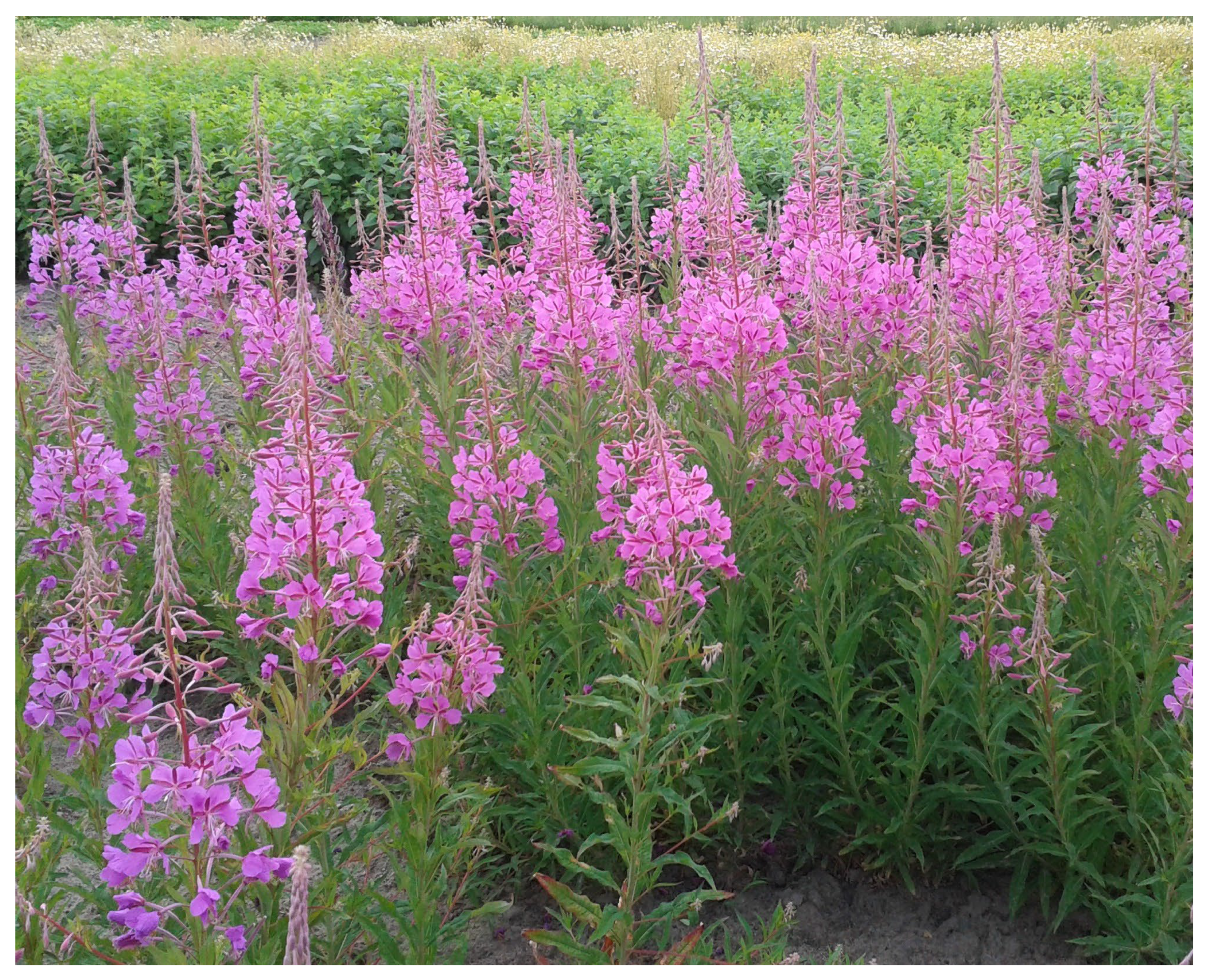
Figure 1.
Epilobium angustifolium during blooming phase.


Table 1.
Studies testing antimicrobial activity of Epilobium angustifolium extracts.
| No. | Plant Material/ Extractant | Experimental Method | Microbial Species | Effect | Reference |
|---|---|---|---|---|---|
| 1 | Roots/EtOH, H2O-EtOH (1:1) | Disk diffusion method | Aspergillus fumigatus, Candida albicans, Cryptococcus neoformans, Microsporum gypseum, Saccharomyces cerevisiae, Trichophyton mentagrophytes | Great antifungal activity against M. gypseum, T. mentagrophytes, S. cerevisiae, and C. albicans. The effect comparable to or even stronger than the positive control (berberine). | [57] |
| 2 | Aerial parts/80% MeOH | Cylinder diffusion method | Aspergillus niger, Candida albicans, Escherichia coli, Staphylococcus aureus | Significant antibacterial effect against E. coli and S. aureus. Slight or lack of antifungal activity against C. albicans and A. niger. | [58] |
| 3 | Aerial parts EtOH | Broth microdilution method (MIC, MCC) | Bacillus subtilis, Candida albicans, C. glabrata, C. krusei, Enterococcus faecalis, Escherichia coli, Klebsiella pneumoniae, Listeria monocytogenes, Microsporum canis, M. gypseum, Pseudomonas aeruginosa, Salmonella enteritidis, Shigella flexneri, Staphylococcus aureus, Streptococcus pyogenes, S. sanguis, Trichophyton mentagrophytes, T. rubrum | Very strong antifungal activity against Microsporum canis and strong antibacterial effect against K. pneumoniae. | [59] |
| 4 | Leaves, flowers, flowering aerial parts/H2O-EtOH (80:20) | Disk diffusion method | Candida albicans, Escherichia coli, Pseudomonas aeruginosa, Staphylococcus aureus | Activity of leaf and flowering part extracts against all microorganisms tested. The strongest effect against C. albicans and S. aureus. | [60] |
| 5 | Seeds/50% MeOH | Disk diffusion method | Candida albicans, Escherichia coli, Pseudomonas aeruginosa, Staphylococcus aureus | Poor activity only against S. aureus. | [61] |
| 6 | Roots/H2O | Broth microdilution method (MIC) | Aspergillus flavus, A. fumigatus, Candida albicans, C. glabrata, C. krusei, C. lusitaniae, C. parapsilosis, C. tropicalis, Cryptococcus neoformans, Epidermophyton floccosum, Fusarium solani, Microsporum canis, Rhizopus sp., Saccharomyces cerevisiae, Trichophyton mentagrophytes, T. rubrum, T. tonsurans | Strong antifungal effect, especially against C. glabrata, C. lusitaniae, and S. cerevisiae strains. | [62] |
| 7 | Whole plant/crude extract (Fytokem, SK, Canada) | Broth microdilution method (absorbance measurement) | Escherichia coli, Micrococcus luteus, Pseudomonas aeruginosa, Staphylococcus aureus | Activity against all bacteria tested. More effective than tetracycline against Gram-negative bacteria: E. coli and P. aeruginosa. | [63] |
| 8 | Leaves, flowers without stems/96% EtOH | Disk diffusion method, Broth microdilution method (MIC), Cell viability test (C. albicans) | Bacillus subtilis, Candida albicans, C. dubliniensis, C. tropicalis, Escherichia coli, Proteus mirabilis, Pseudomonas aeruginosa, Saccharomyces cerevisiae, Staphylococcus aureus | Weak antimicrobial activity, usually similar for both types of extracts (leaves and flowers). | [64] |
| 9 | Aerial parts/75% MeOH | Disk diffusion method, Quorum sensing (QS) inhibition activity assay | Chromobacterium violaceum | Dose-dependent QS inhibition activity. Reduction of violacein production by 41% at 250 μg/mL and 57% at 500 μg/mL concentration of plant extract. | [65] |
| 10 | Flowering aerial parts/EtOH, MeOH | Well diffusion method, Broth microdilution method (MIC, MBC) | Bacillus cereus, B. subtilis, Candida albicans, C. krusei, C. tropicalis, Enterococcus faecalis, E. hirae, Escherichia coli, Klebsiella pneumoniae, Pseudomonas aeruginosa, Proteus vulgaris, Salmonella Typhimurium, Staphylococcus aureus | Moderate antibacterial activity against B. cereus and B. subtilis, low—against S. aureus and P. vulgaris. No antifungal effect against Candia spp. | [13] |
| 11 | Local market, Turkey/99% EtOH, H2O, 95% HE, 99.7% MeOH | Disk diffusion method | Escherichia coli, Klebsiella pneumoniae, Pseudomonas aeruginosa, Staphylococcus aureus | Broad differentiation between antibacterial activity of particular extracts. The greatest sensitivity of P. aeruginosa, K. pneumoniae, and S. aureus to MeOH extract, while E. coli to EtOH and MeOH extracts. | [66] |
| 12 | OMEGA PHARMA Srl (Cantù, Italy)/H2O | Broth microdilution method (MIC) | Arthroderma crocatum, A. currey, A. gypseum, A. insingulare, A. quadrifidum, Bacillus cereus, B. subtilis, Candida albicans, C. parapsilosis, C. tropicalis, Escherichia coli, Pseudomonas aeruginosa, Salmonella Typhi, Staphylococcus aureus, Trichophyton erinacei, T. mentagrophytes, T. rubrum, T. tonsurans | Strong antimicrobial activity, especially against T. tonsurans, T. rubrum, A. crocatum, A. currey, A. insingulare, C. tropicalis. | [67] |
| 13 | Flowering aerial parts/no data | Well diffusion method, Broth microdilution method (MIC, MBC) | Bacillus cereus, Candida spp., Escherichia coli, Staphylococcus aureus | Strong antibacterial activity of 30 and 50% solution of extract against B. cereus and S. aureus. | [68] |
| 14 | Flowering aerial parts/EtOH | Disk diffusion method, Broth microdilution method (MIC) | Escherichia coli, Salmonella Typhimurium, Staphylococcus aureus | Low antibacterial effect compared to ciprofloxacin (disk diffusion method), while significant activity determined by the broth microdilution method. | [69] |
| 15 | Flowering aerial parts/70% EtOH | Well diffusion method | Bacillus pseudomycoides, B. subtilis, B. thuringiensis, Enterococcus faecalis, E. faecium, Pseudomonas aeruginosa, P. fluorescens, Serratia lutea, S. marcescens, Streptococcus pneumoniae | Strong antibacterial activity against S. lutea, S. marcescens, as well as B. pseudomycoides and B. subtilis. | [70] |
| 16 | Aerial parts/H2O, MeOH | Disk diffusion method | Enterobacter cloacae, Escherichia coli, Klebsiella pneumoniae, Proteus vulgaris, Pseudomonas aeruginosa, Salmonella Typhimurium, Serratia marcescens, Staphylococcus aureus, S. epidermidis, Streptococcus pyogenes | Strong antibacterial activity against S. aureus, S. epidermis, S. pyogenes, and P. vulgaris. | [71] |
| 17 | Flowers/H2O | Well diffusion method, Broth microdilution method (MIC, MBC) | Candida albicans, Escherichia coli, Staphylococcus aureus | Strong antimicrobial activity against S. aureus, E. coli, and C. albicans. | [72] |
| 18 | Flowering aerial parts/70% EtOH, 70% iPrOH, H2O in hydrogels | Well diffusion method | Bacillus subtilis, B. pseudomycoides, Enterococcus faecalis, E. faecium, Escherichia coli, Pseudomonas fluorescens, Sarcina lutea, Serratia marcescens, Staphylococcus aureus, Streptococcus epidermidis, S. pneumoniae | Moderate activity against S. pneumoniae, E. faecalis, E. faecium, S. lutea, and E. coli. | [73] |
| 19 | No data/infusion | In vitro digested infusion model. Optical density (OD600) with a Bioscreen C system | Bifidobacterium adolescentis, B. longum, Enterobacter cloacae, Enterococcus faecalis, Escherichia coli, Lactobacillus rhamnosus | No growth inhibition of L. rhamnosus, Bifidobacterium sp. contrary to E. coli. | [74] |
| 20 | Leaves/DCM, MeOH, H2O | Disk diffusion method, Anti-biofilm assay | Bacillus subtilis, Candida maltosa, Escherichia coli (including strains forming biofilms), Pseudomonas aeruginosa, Staphylococcus aureus | Strong antimicrobial activity against C. maltose, significant—against S. aureus, E. coli, P. aeruginosa. Poor or no activity of DCM extract. Weak or moderate anti-biofilm activity. | [75] |
| 21 | Leaves/H2O | Broth microdilution method (MIC) | Bacillus cereus, B. subtilis, Clostridium sporogenes, Enterococcus faecalis, Escherichia coli, Lactobacillus rhamnosus, L. plantarum, Listeria innocua, L. monocytogenes, Salmonella Hofit, S. enterica, Staphylococcus aureus | Significant activity against S. aureus, L. monocytogenes, Bacillus spp., Lactobacillus spp. No activity against Gram-negative bacteria. Resistance of P. aeruginosa. | [56] |
| 22 | Leaves, flowering aerial parts/30% EtOH | Disk diffusion method, Broth microdilution method (MIC) | Bacillus cereus, Candida albicans, Enterococcus faecalis, Escherichia coli, Listeria monocytogenes, Pseudomonas aeruginosa, Salmonella enteritidis, Staphylococcus aureus | Moderate activity against B. cereus and E. faecalis. No activity against Gram-negative bacteria. | [76] |
| 23 | Aerial parts/H2O/silver nitrate nanoparticles | Well diffusion method, Broth microdilution method (MIC) | Acinetobacter baumannii, Escherichia coli, Klebsiella pneumoniae, Pseudomonas aeruginosa, Staphylococcus aureus (MRSA) | Strong antibacterial effect against MRSA. Significant activity against A. baumannii in well-diffusion assay. | [55] |
Abbreviations: DCM—dichloromethane; EtOH—ethanol; HE—n-hexane; MeOH—methanol; iPrOH -isopropanol; MCC—minimum cytocidal concentration; MIC—minimum inhibitory concentration; MDR—multi-drug-resistant; MRSA—methicillin-resistant Staphylococcus aureus.
Table 2.
Antibacterial activity of Epilobium angustifolium extracts.
| Genus | Species | Strains | Inhibition Zone | MIC/OD |
|---|---|---|---|---|
| Bacillus | cereus | PeruMycA 4 | 62.5–125 µg/mL [67] | |
| NCTC 74 | 17.0–25.0 mm [68] | 30% solution [68] | ||
| ATCC 11778 | 13.8–14.1 mm [76] | 0.25% solution [56], MIC50 = 1/64 (aerial parts), MIC50 = 1/128 (leaves) [76] | ||
| NRRL B-3711 | activity found; no data [13] | 1.56 mg/mL [13] | ||
| pseudomycoides | not specified | 6.0–11.5 mm [70], 10.0–13.0 mm [73] | ||
| subtilis | ATCC 6633 | 13.5 mm [64], activity found; no data [13] | 325 µg/mL [59], 1.56 mg/mL [13], 4.6–6.1 mg/mL [64], 0.25% solution [56] | |
| PeruMycA 6 | 125–250 µg/mL [67] | |||
| ATCC 6059 | no activity [75] | 0.25% solution [56] | ||
| not specified | 6.5–11.0 mm [70], 6.5–8.5 mm [73] | |||
| thuringiensis | not specified | 5.5–9.0 mm [70] | ||
| Clostridium | sporogenes | ATCC 11437 | no activity at 0.5% solution [56] | |
| Bifidobacterium | adolescentis | DSM 20083 | no activity (OD) [74] | |
| longum | DSM 20088 | no activity (OD) [74] | ||
| Enterococcus | faecalis | ATCC 29212 | 16.0–17.0 mm [73], 7.9–8.4 mm [76], 5.0–7.0 mm [70], no activity [13] | MIC50 = 1/64 (aerial parts), MIC50= 1/128 (leaves) [76] |
| ATCC 51229 | no activity 0.5% solution [56] | |||
| JCM 1513 | stimulation of growth (OD) [74] | |||
| clinically isolated strain | no activity [59] | |||
| faecium | not specified | 14.5–16.0 mm [73], 5.0–7.0 mm [70] | ||
| hirae | ATCC 9790 | no activity [13] | ||
| Lactobacillus | rhamnosus | B-445, ŁOCK 0900 | no activity 0.5% solution [56], no activity (OD) [74] | |
| plantarum | 299v | no activity 0.5% solution [56] | ||
| Listeria | innocua | ATCC 33090 | 0.25% solution [56] | |
| monocytogenes | ATCC 19111 | 0.25% solution [56], no activity [59] | ||
| ATCC 13932 | 8.2–8.8 mm [76] | MIC50 = 1/64 (aerial parts), MIC50 = 1/128 (leaves) [76] | ||
| ATCC 15313 | no activity 0.5% solution [56] | |||
| ATCC 7644 | no activity 0.5% solution [56] | |||
| IFM 1011 | no activity 0.5% solution [56] | |||
| Micrococcus | luteus | clinically isolated strain | activity similar to vancomycin [63] | |
| Staphylococcus | aureus | DSM 20231 | 4.0–10.0 mm [58] | |
| ATCC 12600 | 11.0–17.0 mm [60], 7.0 mm [61] | |||
| ATCC 6538P | 8.8–10.7 mm [76], 13.5–14.0 mm [64] | 325 µg/mL [59], 6.1 mg/mL [64]; MIC50 = 1/64 (aerial parts), MIC50 = 1/128 (leaves) [76] | ||
| ATCC 6538 | 24.0 mm [72], 17.5–20.0 mm [68], 10.0 mm (MeOH) [75], 14.0 mm (H2O) [75] | 0.5 mg/mL [72], 125–250 µg/mL [67], 30% solution [68] | ||
| 4.4 | 0.15% solution [56] | |||
| ATCC 6535 | 0.15% solution [56] | |||
| ATCC 4538 | 0.15% solution [56] | |||
| MFBF 124 (MRSA) | 14.5 mm [64] | 7.6 mg/mL [64] | ||
| ATCC 25923 | 5.0–21.5 mm [66], 13.3–15.0 mm [71], 7.0 mm [69], activity found; no data [13] | 3.13–6.25 mg/mL [13], 156 µg/mL [69], 0.15% solution [56] | ||
| clinically isolated strains | 325 µg/mL [59], less active than vancomycin [63] | |||
| MRSA clinically isolated strain | 21.0 mm [55] | 0.625 µg/mL [55] | ||
| not specified | 6.0–7.0 mm [73] | |||
| epidermidis | ATCC 12228 | 18.2–18.4mm [71] | ||
| Streptococcus | epidermidis | not specified | 6.0–8.5 mm [73] | |
| pneumoniae | ATCC 49619 | 16.5–18.0 mm [73], 5.0–7.0 mm [70] | ||
| pyogenes | ATCC 12345 | 325 µg/mL [59] | ||
| ATCC 19615 | 14.0–16.4 [71] | - | ||
| sanguis | CDC SS 910 | 325 µg/mL [59] | ||
| Acinetobacter | baumannii | clinically isolated MDR 210 | 10.0 mm [55] | 1.25 µg/mL [55] |
| clinically isolated MDR 211 | 14.0 mm [55] | 1.25 µg/mL [55] | ||
| Chromobacterium | violaceum | CV12472 | no activity (for QS inhibition) [65] | |
| Enterobacter | cloacae | ATCC 23355 | no activity [71] | |
| PCM 533 | no activity [74] | |||
| Escherichia | coli | ATCC 8739 | 4.0–10.0 mm [58] | |
| ATCC 15221 | no activity [59] | |||
| ATCC 8677 | 6.0–7.0 mm [60], no activity [60,61] | |||
| ATCC 10535 | 13.5–14.5 mm [64] | 6.1–7.6 mg/mL [64] | ||
| ATCC 10536 | no activity [76] | 62.5–125 µg/mL [67], no activity 0.5% solution [56] | ||
| ATCC 11229 | (H2O) 11.0 mm [75], (MeOH) 13.0 mm [75] | |||
| ATCC 35218 | no activity [13] | |||
| MFBF P11 (p-fimbriae positive) | 14.5 mm [64] | 9.1–16.2 mg/mL [64] | ||
| ATCC 25922 | 21.0 mm [72], 8.0 mm [69], ≤10 mm [68], 11.5–15.0 mm [66], no activity [13,71] | 156 µg/mL [69], 1.0 mg/mL [72], no activity for 30 and 50% solution [68] | ||
| ATCC 25923 | no activity 0.5% solution [56] | |||
| PeruMycA 2 | 125–250 µg/mL [67] | |||
| PeruMycA 3 | 125–250 µg/mL [67] | |||
| PBio 729 (MRGN) | no activity [75] | (MeOH) 50 µg/mL, (H2O) 100 µg/mL [75] | ||
| PBio 730 (MRGN) | no activity [75] | (MeOH) 50 µg/mL, (H2O) 100 µg/mL [75] | ||
| ATCC 11239 | 11.0–13.0 mm [75] | |||
| clinically isolated MDR 55 | 16.0 mm [55] | 0.625 µg/mL [55] | ||
| clinically isolated strain | more active than tetracycline [63] | |||
| not specified | 13.0–15.0 mm [73] | 64 µg/mL [74] | ||
| Klebsiella | pneumoniae | ATCC 10031 | 81 µg/mL [59] | |
| ATCC 13883 | no activity [13,71] | |||
| MDR 104 | 14.0 mm [55] | 0.625 µg/mL [55] | ||
| MMLRD not specified | 13.0–23.5 mm [66] | |||
| Proteus | mirabilis | MFBF 624 | no activity [64] | 15.1–16.2 mg/mL [64] |
| vulgaris | RSKK 96029 | activity found; no data [13] | 3.13 mg/mL [13] | |
| ATCC 13315 | 15.2–17.3 mm [71] | |||
| Pseudomonas | aeruginosa | ATCC 27853 | 10.5–11.5 mm [64], 0.0–12.0 mm [75], no activity [13,71,76] | 162 µg/mL [59], 9.1 mg/mL [64] no activity [71] |
| ATCC 9721 | 7.0 mm [60], no activity [60,61] | |||
| PeruMycA 5 | 62.5–125 µg/mL [67] | |||
| ATCC 2753 | 4.0–6.0 mm [70] | |||
| clinically isolated MDR 40 | 9.0 mm [55] | 1.25 µg/mL [55] | ||
| clinically isolated MDR 215 | 11.0 mm [55] | 1.25 µg/mL [55] | ||
| MMLRD not specified | 4.0–25.5 mm [66] | |||
| clinically isolated strain | more active than tetracycline [63] | |||
| fluorescens | not specified | 6.0 mm [70], 7.0–8.5 mm [73] | ||
| Salmonella | enterica | ATCC 29631 | 0.15% solution [56] | |
| enteritidis | IAL 1132 | no activity [59] | ||
| ATCC 13076 | no activity [76] | |||
| Hofit | IFM 2318 | no activity 0.5% solution [56] | ||
| Typhimurium | ATCC 14028 | 6.0 mm [69], no activity [13,71] | 312 µg/mL [69] | |
| Typhi | PeruMycA 7 | 125–250 µg/mL [67] | ||
| Sarcina | lutea | ATCC 9341 | 15.0–16.5 mm [73] | |
| Serratia | lutea | ATCC 9341 | 8.0–16.0 mm [70] | |
| marcescens | ATCC 8100 | no activity [71] | ||
| not specified | 7.0–15.0 mm [70], 9.0–10.5 mm [73] | |||
| Shigella | flexneri | CDC 9767 | no activity [59] | |
| IAL 1517 | no activity [59] |
Abbreviations: ATCC—American Type Culture Collection (USA); NRRL—Agricultural Research Service Culture Collection (USA); NCM—National Centre for Mycology in Edmonton (Canada); DSM: Deutsche SammLung von Microorganismen (Germany); FOMK—Division of Pharmacognosy, University of Helsinki (Finland); YMBL—Division of General Microbiology, University of Helsinki (Finland); OMH—Ontario Ministry of Health, Mycology Laboratory, Toronto, (Canada); OGH—Ottawa General Hospital, Microbiology Laboratory, (Canada); MFBF—collection of the Department of Microbiology, Faculty of Pharmacy and Biochemistry, University of Zagreb (Croatia); CMBC—China General Microbiological Culture Collection Center, Beijing (China); MMLRD—Medical Microbiology Laboratory, Research Hospital of Dicle University in Diyarbakir (Turkey); PeruMycA—culture collection of the Department of Chemistry, Biology and Biotechnology, University of Perugia (Italy); CDC—Collecione de Coltura, IAL- Istituto Alberto Luz (Italy); NCTC—The National Collection of Type Cultures, UK Health Security Agency, (UK). MIC—minimum inhibitory concentration; MBC—minimum bactericidal concentration; MDR—multi-drug resistance, MRSA—methicillin-resistant S. aureus; MRGN—Multi-resistant Gram-negative bacilli.
Table 3.
Antifungal activity of Epilobium angustifolium extracts.
| Genus | Species | Strains | Inhibition Zone | MIC |
|---|---|---|---|---|
| Arthroderma | crocatum | CCF 5300 | 15.62–31.25 µg/mL [67] | |
| currey | CCF 5207 | 31.25–62.5 µg/mL [67] | ||
| gypseum | CCF 6261 | 125–250 µg/mL [67] | ||
| insingulare | CCF 5417 | 31.25–62.5 µg/mL [67] | ||
| quadrifidum | CCF 5792 | 62.5–125 µg/mL [67] | ||
| Epidermophyton | floccosum | NCM 335 | - | 3 mg/mL [62] |
| clinically isolated strain | 3 mg/mL [62] | |||
| Microsporum | canis | NCM 336 | 1 mg/mL [62] | |
| clinically isolated strains | 10 µg/mL [59], no growth [62] | |||
| gypseum | OMH FR323 | 18.9 mm [57] | ||
| clinically isolated strains | 650 µg/mL [59], no growth [62] | |||
| Trichophyton | erinacei | CCF 5930 | 125–250 µg/mL [67] | |
| rubrum | CCF 4879 | 62.5–125 µg/mL [67] | ||
| CCF 4933 | 15.62–31.25 µg/mL [67] | |||
| clinically isolated strains | 650 µg/mL [59], no growth, 3000 mg/mL [62] | |||
| mentagrophytes | OMH T2379 | 16.1 mm [57] | ||
| CCF 4823 | 62.25–125 µg/mL [67] | |||
| clinically isolated strains | 162–650 µg/mL [59], 3–6 mg/mL [62] | |||
| tonsurans | NCM 334 | 3 mg/mL [62] | ||
| CCF 4834 | 7.81–15.62 µg/mL [67] | |||
| clinically isolated strain | no growth [62] | |||
| Aspergillus | flavus | clinically isolated strains | no activity [62] | |
| fumigatus | OMH FR2837 | no activity [57] | ||
| NCM 338 | no activity [62] | |||
| clinically isolated strains | no activity [62] | |||
| niger | ATCC 16404 | no activity [58] | ||
| Candida | albicans | ATCC 10231 | 15.0–20.0 mm [60], 16.0 mm [72], 10.0–11.7 mm [76], 1.0–3.0 mm [58], no activity [13,61,64] | 6.1–12.1 mg/mL [64], 4 mg/mL [72] MIC50 = 1/64 (leaves), MIC50 = 1/32 [76] |
| ATCC 24433 | 0.8 mg/mL [62] | |||
| ATCC 90028 | 0.4 mg/mL [62] | |||
| Candida | albicans | ATCC 885–653 | 10.0–15.0 mm [68] | |
| OGH 308–1329 | 13.5 mm [57] | |||
| DBVPG 6379 | 125–250 µg/mL [67] | |||
| DBVPG 6183 | 125–250 µg/mL [67] | |||
| clinically isolated strains | 0.2–1.6 mg/mL [62], 325 µg/mL [59] | |||
| dubliniensis | MFBF 501 | no activity [64] | 12.1 mg/mL [64] | |
| glabrata | clinically isolated strains | 25–50 mg/mL [62], no activity [59] | ||
| krusei | ATCC 6258 | no activity [13] | 0.2 mg/mL [62] | |
| clinically isolated strains | 0.2 mg/mL [62], 325 µg/mL [59] | |||
| maltosa | SBUG 700 | 18.0 mm (H2O), 25 mm (MeOH), no activity (DCM) [75] | ||
| lusitaniae | clinically isolated strains | 0.05–0.2 mg/mL [62] | ||
| parapsilosis | ATCC 90018 | 0.8 mg/mL [62] | ||
| ATCC 22019 | 0.1 mg/mL [62] | |||
| DBVPG 6551 | ≥250 µg/mL [67] | |||
| clinically isolated strains | 0.1–0.4 mg/mL [62] | |||
| tropicalis | ATCC 750 | no activity [64] | 1.6 mg/mL [62], 15.2 mg/mL [64] | |
| Y-12968 | no activity [13] | |||
| DBVPG 6184 | 31.25–62.5 µg/mL [67] | |||
| clinically isolated strain | 1.6 mg/mL [62] | |||
| spp. | ATCC 885–653 | ≤10.0–15.0 mm [68] | 30% solution [68] | |
| Saccharomyces | cerevisiae | ATCC 48252 | 15.5 mm [57] | |
| NCYC 87 | no activity [64] | 18.2 mg/mL [64] | ||
| clinically isolated strains | 0.05 mg/mL [62] | |||
| Cryptococcus | neoformans | OMH FR2704 | 11.5 mm [57] | |
| clinically isolated strains | 0.4 mg/mL [62] | |||
| Fusarium | solani | Pig pasture soil | no activity [62] | |
| Rhizopus | sp. | NCM 337 | 25 mg/mL [62] | |
| clinically isolated strain | 0.2 mg/mL [62] |
Abbreviations: ATCC—American Type Culture Collection, Rockville (USA); NRRL—Agricultural Research Service Culture Collection (USA); NCM—National Centre for Mycology in Edmonton (Canada); CCF—Culture Collection of Fungi, Department of Botany, Charles University, Prague (Czech Republic); DSM—Deutsche Sammlung von Microorganismen (Germany); OMH—Ontario Ministry of Health, Mycology Laboratory, Toronto (Canada); OGH—Ottawa General Hospital, Microbiology Laboratory (Canada); MFBF—collection of the Department of Microbiology, Faculty of Pharmacy and Biochemistry, University of Zagreb (Croatia); SBUG—Department of Biology, University of Greifswald (Germany); NCYC—National Collection of Yeast Cultures (UK); H2O—aqueous extract; MeOH—methanol extract; DCM—dichloromethane extract.
Disclaimer/Publisher’s Note: The statements, opinions and data contained in all publications are solely those of the individual author(s) and contributor(s) and not of MDPI and/or the editor(s). MDPI and/or the editor(s) disclaim responsibility for any injury to people or property resulting from any ideas, methods, instructions or products referred to in the content. |
© 2023 by the authors. Licensee MDPI, Basel, Switzerland. This article is an open access article distributed under the terms and conditions of the Creative Commons Attribution (CC BY) license (https://creativecommons.org/licenses/by/4.0/).
Share and Cite
MDPI and ACS Style
Dreger, M.; Adamczak, A.; Foksowicz-Flaczyk, J. Antibacterial and Antimycotic Activity of Epilobium angustifolium L. Extracts: A Review. Pharmaceuticals 2023, 16, 1419. https://doi.org/10.3390/ph16101419
AMA Style
Dreger M, Adamczak A, Foksowicz-Flaczyk J. Antibacterial and Antimycotic Activity of Epilobium angustifolium L. Extracts: A Review. Pharmaceuticals. 2023; 16(10):1419. https://doi.org/10.3390/ph16101419
Chicago/Turabian StyleDreger, Mariola, Artur Adamczak, and Joanna Foksowicz-Flaczyk. 2023. "Antibacterial and Antimycotic Activity of Epilobium angustifolium L. Extracts: A Review" Pharmaceuticals 16, no. 10: 1419. https://doi.org/10.3390/ph16101419
Note that from the first issue of 2016, this journal uses article numbers instead of page numbers. See further details here.