
Enlarge
Donor: Royal Victoria Hospital
Date: 1933
Size (H x W x D cm): 9 x 9 x 4
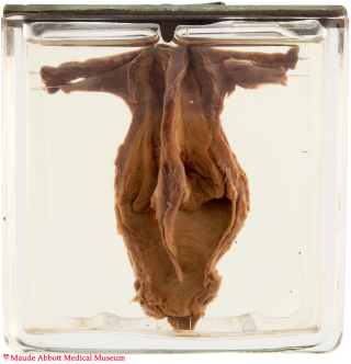
Two completely separate uterine cavities, cervices and vaginas are present.
History: Stillborn female.
Comment: The uterus and upper portion of the vagina are derived from the Müllerian ducts, which arise in the urogenital ridge during the 5th to 6th week of development as longitudinal invaginations of the coelomic epithelium. In subsequent weeks, the two ducts approach each other in the midline, eventually fusing to form the uterovaginal primordium (UVP). Fusion and canalization of the portion of the UVP destined to be the uterus normally results in a single chamber. However, abnormalities in this process can result in a variety of anatomic variants including uterus didelphys and bicornuate uterus (see specimens 28 and 29). The vagina is believed to develop by fusion of the lower portion of the UVP (forming its upper part) with a portion of the urogenital sinus (forming the inferior part). Initially solid, this fused structure normally undergoes vacuolization to produce a single cavity.
Müllerian duct abnormalities are relatively common, the prevalence estimated to be about 2 - 4% in the general population and up to 10% in women with recurrent pregnancy loss. However, pronounced abnormalities such as illustrated here are uncommon. Most anomalies are sporadic; however, associations with a number of genetic syndromes are described.
AFS Classification of Anomalies of the Müllerian Duct
(Adapted from the American Fertility Society classifications of adnexal adhesions, distal tubal occlusion, tubal occlusion secondary to tubal ligation, tubal pregnancies, Müllerian anomalies and intrauterine adhesions. Fertil Steril. Jun 1988;49(6):944-55.)
|
Classification |
Clinical Finding |
Description |
|
I |
Segmental or complete agenesis or hypoplasia |
Agenesis and hypoplasia may involve the vagina, cervix, fundus, tubes, or any combination of these structures. |
|
II |
Unicornuate uterus with or without a rudimentary horn |
When an associated horn is present, this class is subdivided into communicating (continuity with the main uterine cavity) and noncommunicating (no continuity with the main uterine cavity). |
|
III |
Didelphys uterus |
Complete or partial duplication of the vagina, cervix, and uterus. |
|
IV |
Complete or partial bicornuate uterus |
Complete bicornuate uterus is characterized by a uterine septum that extends from the fundus to the cervical os. The partial bicornuate uterus demonstrates a septum, which is located at the fundus. In both variants, the vagina and cervix each have a single chamber. |
|
V |
Complete or partial septate uterus |
A complete or partial midline septum is present within a single uterus. |
|
VI |
Arcuate uterus |
A small septate indentation is present at the fundus. |
|
VII |
DES-related abnormalities |
A T -shaped uterine cavity with or without dilated horns. |