PHONE
+44-7482-878921
+44-7482-878921
2376-0249
Case Blog - International Journal of Clinical & Medical Images (2014) Volume 1, Issue 2

Author(s): Ujwala Shivarama Shetty*
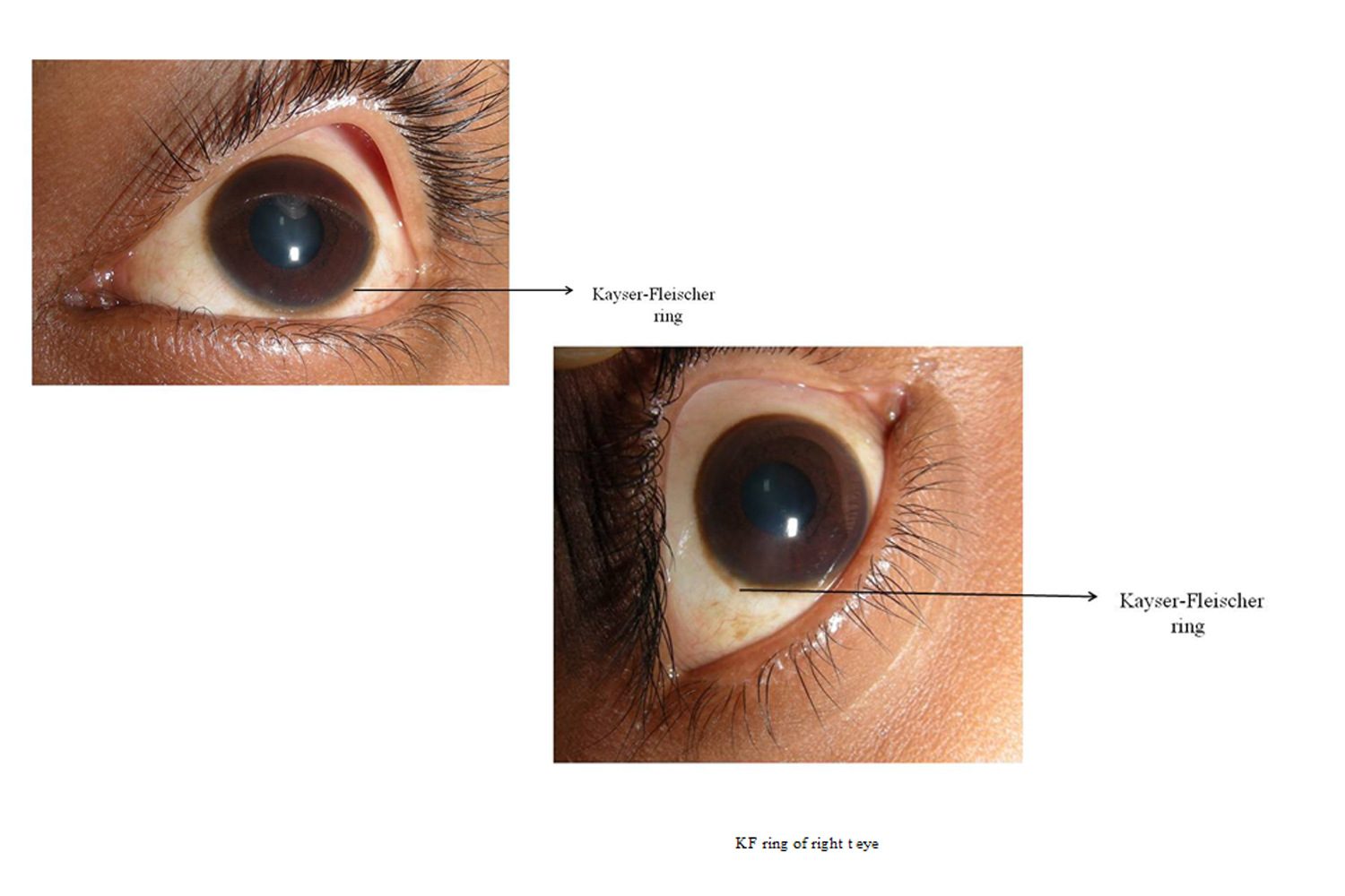
In this paper we present a case of 16 years old female patient who came to our department with pain in the tooth since three days. Intra oral examination revealed grossly decayed tooth. On general physical examination a dark ring was seen encircling the iris of both right and left eye (Kayser-Fleischer ring- KF ring, Figure 1 and 2). Lab investigations report revealed serum copper – 114mg/dl, urinary copper – 30mg/dl and cerulloplasmin – 20mg/dl. MRI revealed abnormal signal in basal ganglion.Patient was diagnosed as having Wilson’s disease and is being treated with Penicillamine, Pictane and Zinc medications for life.
Wilson\'s disease is also known as Hepatolenticular degeneration. It is an autosomal-recessive disorder. In this condition there is disturbance of copper metabolism which affects the liver. This in turn result is abnormal deposition of copper, initially in the hepatocytes and later in various organs and tissues, particularly the CNS, the corneas, and the kidneys [3]. The prevalence of this disease is 1 in 30,000 [4]. The patients usually present first time as adolescents or young adults. The incidence is somewhat more common in Indian children.5 It is caused by impaired function of P-type adenosine triphosphatase (ATPase), encoded by ATP7B gene located on chromosome 13q14 and consists of 21 exons [2].
Diagnostic features of Wilson’s disease
1) Decreased levels of Serum cerulloplasmin.
2) K.F. ring (Kayser-Fleischer ring)
3) Liver biopsy to estimate hepatic copper contents [5].
Kayser-Fleischer rings are due to deposition of copper within Descemet’s membrane. Kayser-Fleischer rings are most of the time always bilateral, but unilateral formation has also been reported. The colour of the rings range from gold to brown to green. Ring formation of ring first becomes visible in the superior aspect of the cornea, which is then followed by the inferior aspect, with subsequent filling in of the medial and lateral aspects. The pigment appears first in the corneal periphery at the limbus and then spreads centrally [6].
Diagnosis of Wilson’s disease is mainly based on the clinical and laboratory findings. The main goal of the treatment is to reduce the amount of copper in the liver and other tissues by administering copper chelating agents and low copper diet [4].
Treatment
1. Restriction of food containing copper like fish, nuts, chocolates etc.
2. An oral chelating agent- D-Penicillamine 20 mg/kg body weight.
3. Zinc supplements 25 mg thrice a day which reduces copper absorption from gut.
4. Vitamin B6 is given to counteract the antifolate effect of D-Penicillamine [5].
To conclude in this paper we report a case of Wilson’s disease who had KF ring which is one of the diagnostic features of this disease. Though Wilson’s disease is a rare entity its diagnosis at early stage is important so that further neurologic and hepatic damage can be prevented.
References
Corresponding author
Ujwala Shivarama Shetty
Department of Opthalmology
India
 Awards Nomination