
Gynecological Pathology Image Atlas
High grade squamous intraepithelial lesion: Cervix
Overview
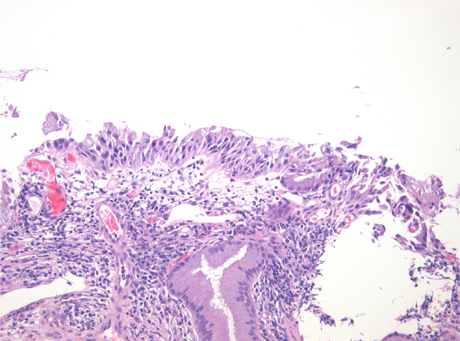
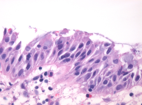
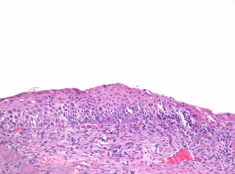
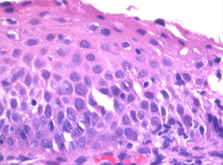
High grade squamous intraepithelial lesion (HSIL) of the cervix is a pre_invasive lesion caused by oncogenic types of human papillomavirus (HPV), the most common types being viral type 16 and type 18. On histology, HSIL shows atypical cells with irregular nuclear contour, increased nuclear size, hyperchromasia, coarse chromatin, increased mitotic activity and increased nuclear/cytoplasmic ration. HSIL can show persistence of koilocytic activity on the surface, but it is distinguished from LSIL when these atypical cells with high N/C ratio occupy more then 1/3 of the thickness of the epithelium. Most HSIL are easily recognized (Fig.1) and their diagnosis has excellent inter_observer and intra_observer agreement. However, the diagnosis of HSIL can be particularly challenging when the epithelium is very thin (Fig.2) or when the cells share similar morphology with immature squamous metaplasia (Fig.3). In these situations, particular attention should be given to the nuclear features, the presence of polymorphism, mitotic activity and coarse chromatin. In many cases, immunostaining with p16 and ki67 can be very helpful to confirm the diagnosis of a HSIL.
Click the thumnails to enlarge image gallery