Meconium Ileus Uncomplicated

  • Etiology: thick meconium causing in-utero distal ileal obstruction
  • AXR: bubbly bowel contents in right lower quadrant
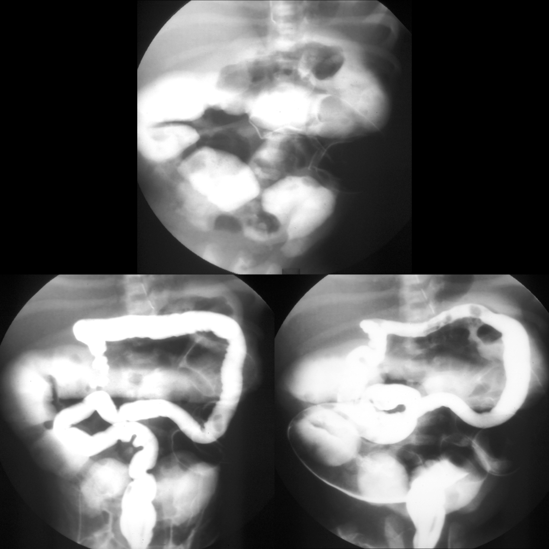
  • Enema: smallest of all microcolons with multiple small meconium filling defects in colon and terminal ileum and normal caliber terminal ileum with a very dilated distal ileum
  • Treatment: high osmolar water soluble contrast enema (diluted 1 part contrast: 2 parts sodium chloride) which must be refluxed above terminal ileum into dilated distal ileum in order to be successful (50% success rate)
  • Clinical: most cases are associated with cystic fibrosis, 20% of cystic fibrosis patients develop it

Radiology Cases of Meconium Ileus Uncomplicated

Enema of meconium ileus
Enema shows on the early AP view (left) a microcolon and on the later lateral view (right) reflux of contrast into a dilated terminal ileum.
Barium enema of uncomplicated meconium ileus
Early (left) image from a water-soluble contrast enema shows a microcolon and reflux of contrast into a non-dilated terminal ileum. Later (right) image from the same enema shows reflux of contrast into dilated loops of ileum, which are above the level of the obstruction by meconium in the terminal ileum.
Enema of successful reduction of uncomplicated meconium ileus
AXR AP (above) obtained after an outside upper GI exam which demonstrated normal bowel rotation shows contrast filling multiple dilated loops of small bowel. Initial AP image from a water soluble enema (below left) shows a microcolon with contrast almost reaching the cecum. Later image from the enema (below right) shows reflux of contrast into dilated loops of distal ileum in the right lower quadrant.