- Etiology: abnormality of neuronal migration characterized by undermigration
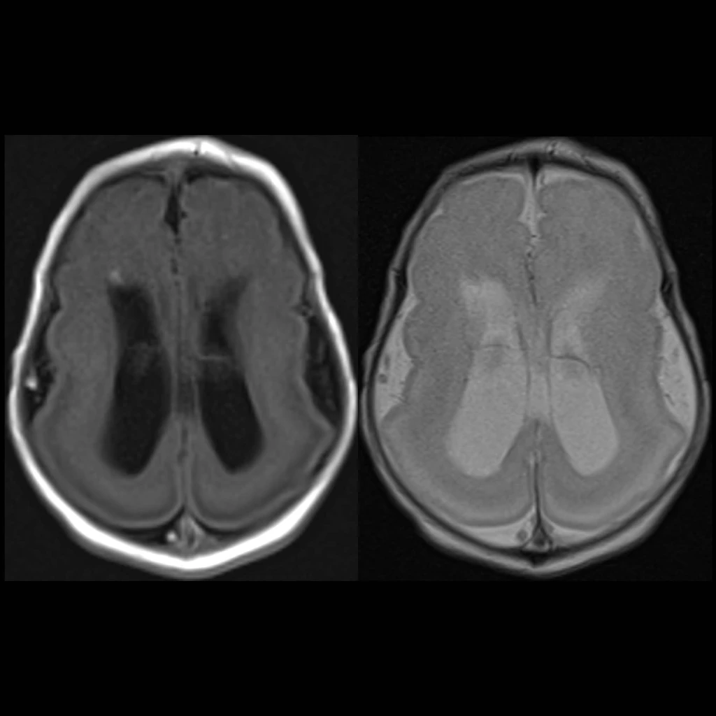
- Imaging: absent gyri giving a smooth brain appearance, shallow vertical Sylvian fissues giving brain an hourglass or figure of 8 appearance on axial images, thickened “cortex” – usually thinner outer layer and thicker inner layer with thin white matter layer separating the two (“cell sparse zone”), diminished cerebral white matter and often callosal hypogenesis, ventriculomegaly, occasionally cerebellar hypoplasia
- DDX:
- Complications:
- Treatment:
- Clinical: developmental delay and seizures, agyria = complete lissencephayly, pachygyria = incomplete lissencephaly, band heterotopia is milder form of classic lissencephaly
Radiology Cases of Lissencephaly Type I Classic