Polymicrogyria

  • Etiology: abnormal cortical organization secondary to ischemic laminar necrosis of 5th cortical layer after 20 weeks gestation, may be caused by in-utero cytomegalovirus, in-utero ischemia, or chromosomal mutation
  • Imaging: usually multiple small gyri, too few sulci, focal or diffuse, unilateral or bilateral, in and around Sylvian fissure, anomalous venous drainage in 50%, sometimes indistinguishable from pachygyria
  • DDX: gyri of polymicrogyria cannot be named by textbook while gyri of lissencephaly can be named by textbook though simplified
  • Complications:
  • Treatment:
  • Clinical:

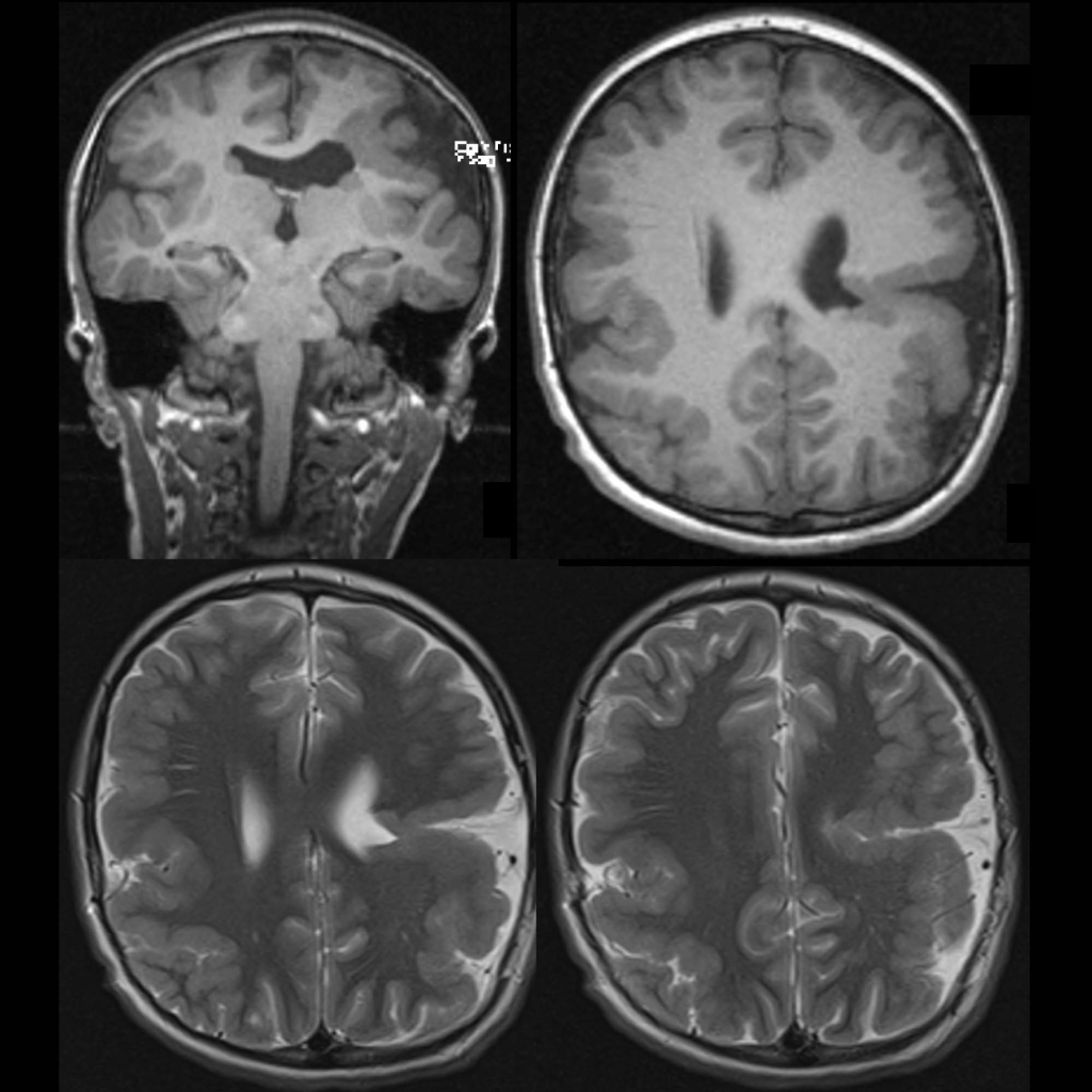
Radiology Cases of Polymicrogyria

MRI of diffuse polymicrogyria in a patient with cytomegalovirus encephalitis
Axial T2 MRI without contrast of the brain show thickening and abnormal sulcation of the perirolandic gray matter diffusely bilaterally. There is also evidence of perisylvian fissure syndrome bilaterally.
MRI of perisylvian polymicrogyria and open lip schizencephaly
Coronal and axial T1 (above) and axial T2 (below) MRI without contrast of the brain shows small haphazard appearing gyri and too few sulci present bilaterally, left greater than right, primarily in the sylvian fissures. There is also a cleft of gray matter with cerebrospinal fluid within extending from the cortex to the posterior aspect of the left lateral ventricle.
MRI of perisylvian polymicrogyria
Axial T2 (above) and coronal T1 (below) MRI without contrast of the brain shows small haphazard appearing gyri and too few sulci present bilaterally, primarily in the sylvian fissures.