Congenital Lobar Emphysema

  • Etiology: intrinsic airway lumen narrowing due to dysplastic bronchial cartilage or extrinsic airway lumen narrowing leading to congenital ball valve effect and hyperinflated lobe
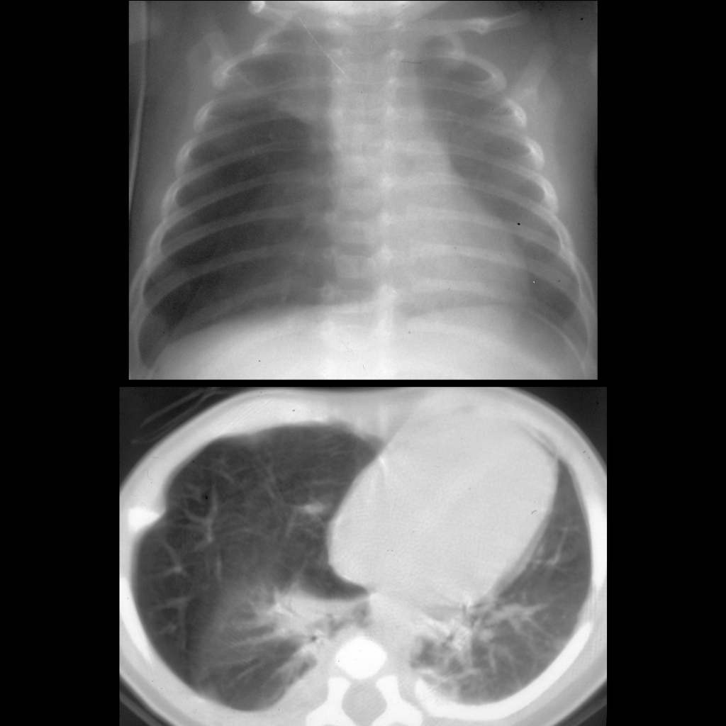
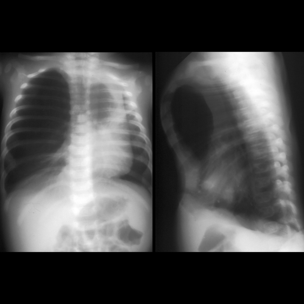
  • CXR: single large lesion with retention of fluid at birth and air trapping later
  • Imaging: Usually unilateral and involves one lobe, usually affects upper lobes (left upper lobe > right middle lobe > right upper lobe > lower lobes), has normal blood supply from pulmonary artery
  • Clinical: symptomatic during infancy

Radiology Cases of Congenital Lobar Emphysema

CXR of congenital lobar emphysema
CXR on day 1 (above) shows a semi-solid mass in the right middle lobe obscuring the right heart border. CXR on day 5 (below) shows the right middle lobe mass has now cleared of fluid and has filled with air and is causing mediastinal shift to the left.
CXR and CT of congenital lobar emphysema
CXR AP (above) and axial CT without contrast of the chest (below) show hyperinflation of the right middle lobe causing mediastinal shift to the left and partial collapse of the right upper and lower lobes.
CXR of congenital lobar emphysema
CXR AP and lateral show overinflation of the right upper lobe that is herniating across the midline.

Clinical Cases of Congenital Lobar Emphysema

Clinical image of congenital lobar emphysema
Clinical image shows a hyperexpanded right hemithorax when compared to the left hemithorax.

Surgery Cases of Congenital Lobar Emphysema

Surgical image of congenital lobar emphysema
Surgical image taken just after opening the right chest shows a very large and emphysematous right upper lobe which was allowed to herniate out through the incision, thus decompressing the other intrathoracic structures. The patient’s respiratory status improved immediately. The tip of the normal right middle lobe can be seen under the over inflated right upper lobe.

Gross Pathology Cases of Congenital Lobar Emphysema

Gross pathological image of congenital lobar emphysema
Gross pathological image shows a distended and emphysematous right upper lobe with focal areas of hemorrhage and subpleural bullae.

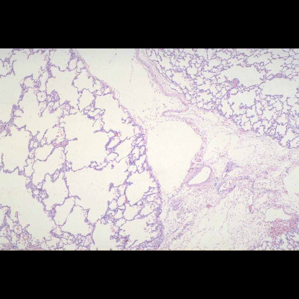
Histopathology Cases of Congenital Lobar Emphysema

Histopathology of congenital lobar emphysema
Histopathological image of H&E stained section shows the dilated but normally formed alveoli (on the left) separated by less expanded alveoli in a small adjacent lobe (on the right).