Pediatric Arachnoid Cyst

  • Etiology: can be primary or secondary, anterior middle cranial fossa most common, left > right, large size range
  • Imaging: circumscribed CSF signal collection, most don’t change substantially over time especially in children > 4 years old
  • DDX:
  • Complications: spontaneous rupture of cysts causing subdural hygroma is uncommmon – contact sports may be restricted with large cysts, intracystic hemorrhage after trauma rare
  • Treatment: indicated if causing clear and specific neurologic symptoms, mass effect alone not indication for surgery
  • Clinical: very common – 2% of scans, usually asymptomatic and incidental

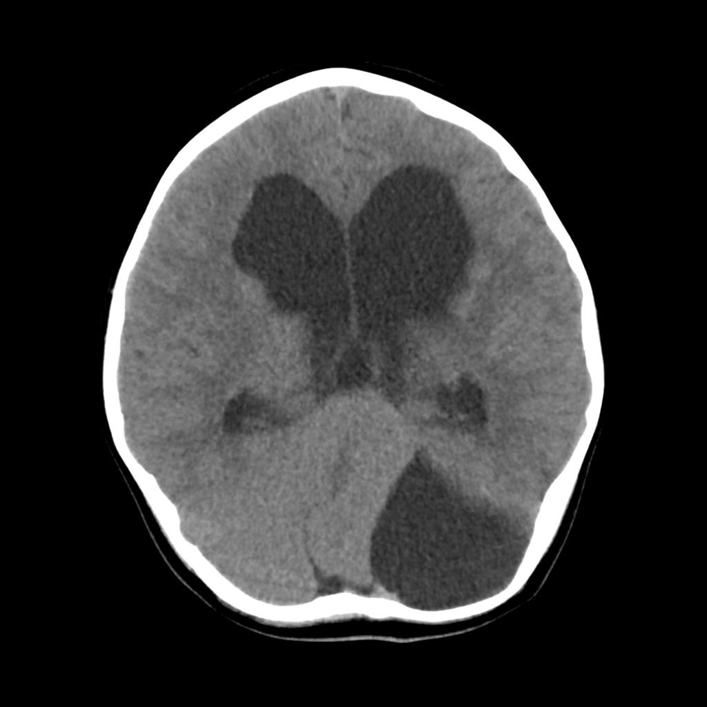
Radiology Cases of Arachnoid Cyst

MRI of hemispheric arachnoid cyst
Axial T2 (left) and coronal T1 (right) MRI without contrast of the brain show a large extra-axial cystic structure in the right hemisphere causing significant mass effect to the left.
CT of arachnoid cyst of the posterior fossa
Axial CT without contrast of the brain shows a cystic structure on the left side of the posterior fossa that is shifting the cerebellum, which is structurally normal, to the right. The fourth ventricle is compressed leading to a moderate amount of hydrocephalus.

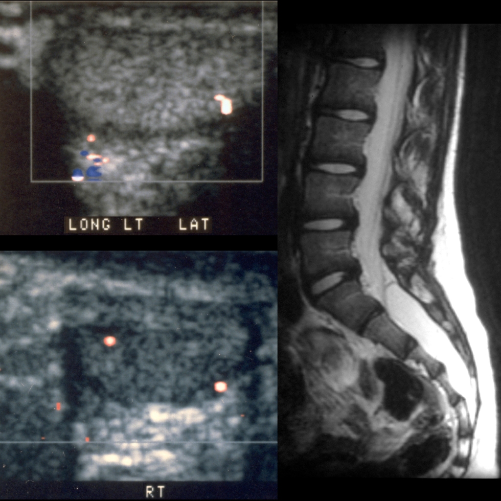
Radiology Cases of Sacral Arachnoid Cyst

MRI of arachnoid cyst of the sacrum
Sagittal color doppler US of the left (above left) and right (below left) testicles shows normal appearing color doppler flow to each testicle which was confirmed on spectral dopper US. Sagittal T2 MRI without contrast of the lumbar spine (right) shows a large cylindrical structure filling the sacral spinal canal from S1-S5 whose signal intensity followed cerebrospinal fluid on all sequences.