Pediatric Arachnoid Cyst of Posterior Fossa

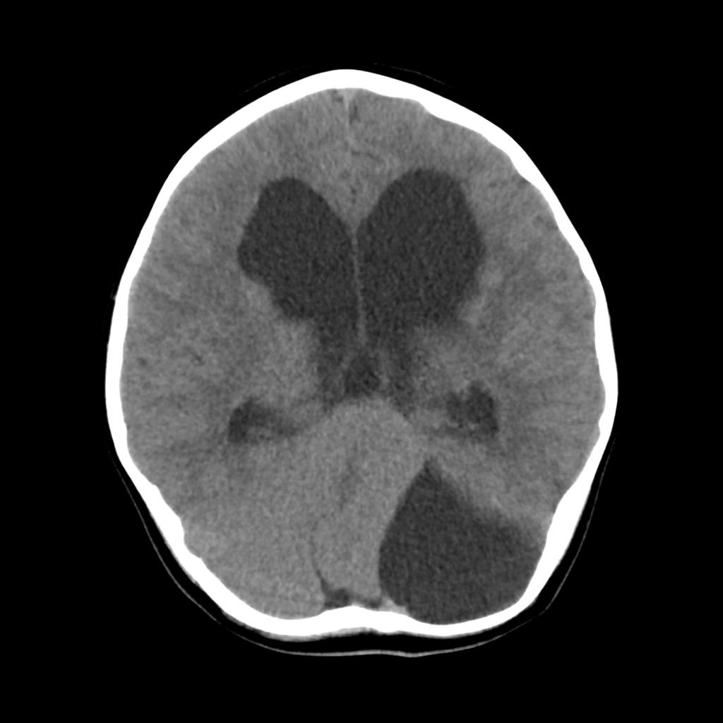
  • Etiology: congenital splitting of arachnoid layer with accumulation of CSF within this potential space
  • Imaging: normal inferior vermis, may elevate torcula, may erode calvarium, cyst may extend to cerebellopontine angle wrapping around cerebellar hemisphere

Radiology Cases of Arachnoid Cyst of Posterior Fossa

CT of arachnoid cyst of the posterior fossa
Axial CT without contrast of the brain shows a cystic structure on the left side of the posterior fossa that is shifting the cerebellum, which is structurally normal, to the right. The fourth ventricle is compressed leading to a moderate amount of hydrocephalus.