Agenesis of Corpus Callosum

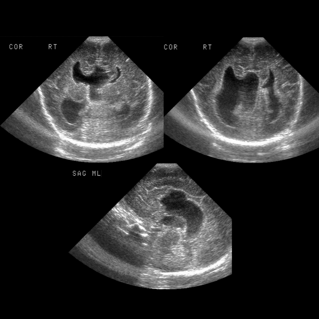
  • Etiology: abnormality of ventral induction, order of growth is genu -> body -> splenium (front to back), Bundles of Probst run front to back instead of crossing midline
  • Imaging: parallel lateral ventricles on axial images, Viking horn / everted lateral ventricles on coronal images, colpocephaly, high riding third ventricle
    — complete: absence all 4 parts of corpus callosum (rostrum, genu, body, splenium)
    — partial: absence of posterior body +/- splenium, short corpus callosum with +/- missing parts and +/- abnormal shape, at least one of segments visible, at least one of segments missing and length reduced, interrupted or short corpus callosum
  • DDX: corpus callosum anomalies
    — agenesis – either complete or partial
    — hypoplasia – characterized by the presence of a fully formed but thinner corpus callosum
    — hyperplasia – characterized by the presence of a fully formed but thick corpus callosum
    — dysplasia defined as a corpus callosum with a hump shape
  • Complications:
  • Treatment:
  • Clinical: associated with pericallosal lipoma which arise from persistence + maldifferentiation of meninx primitiva, Dandy Walker malformation, encephalocele

Radiology Cases of Agenesis of Corpus Callosum

Head US of agenesis of the corpus collosum with large interhemispheric cyst
Coronal (left) and sagittal (right) US of the brain show a Viking horn appearance of the anterior horns of the lateral ventricles and absence of the corpus callosum and a large midline cyst.
Head US of agenesis of the corpus callosum with a small interhemispheric cyst
Coronal US of the brain shows a Viking horn deformity of the anterior horns of the lateral ventricles (above left) and parallel bodies of the lateral ventricles (above right). There is mild hydrocephalus present and the third ventricle is high riding. Sagittal US of the brain (below) shows absence of the corpus callosum.
Head US of agenesis of the corpus callosum
Coronal US of the brain shows a Viking horn appearance of the anterior horns of the lateral ventricles (above left) and parallel bodies of the lateral ventricles (above right). Sagittal US of the brain (below) shows absence of the corpus callosum.
MRI of Chiari II malformation
Sagittal T1 MRI without contrast of the brain shows a small posterior fossa with downward cerebellar tonsil herniation and a small fourth ventricle. There is kinking of the spinal cord at the cervico-medullary junction. There is also agenesis of the body and rostrum of the corpus callosum.