12 October 2022: Case of the Month #520

Thanks to Dr. Debra Zynger, Ohio State University, Columbus, Ohio, USA, for contributing this case and discussion, and to Dr. Bonnie Choy, Northwestern University, Chicago, Illinois, USA, for reviewing the discussion.

Clinical history:

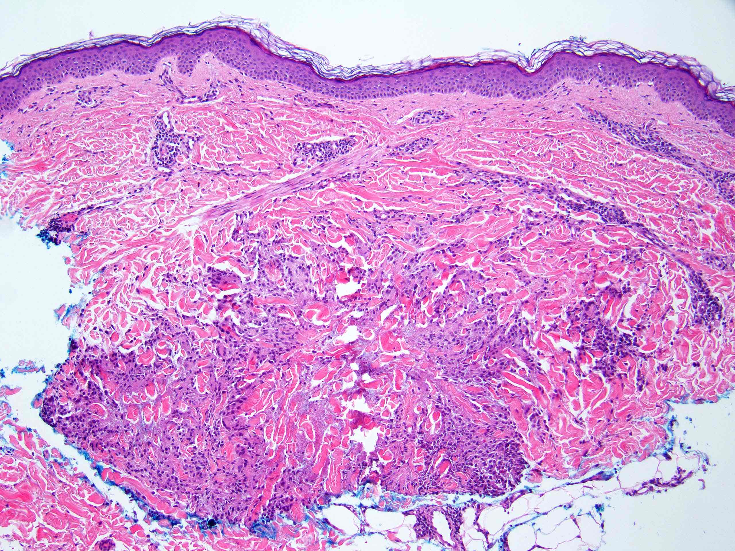
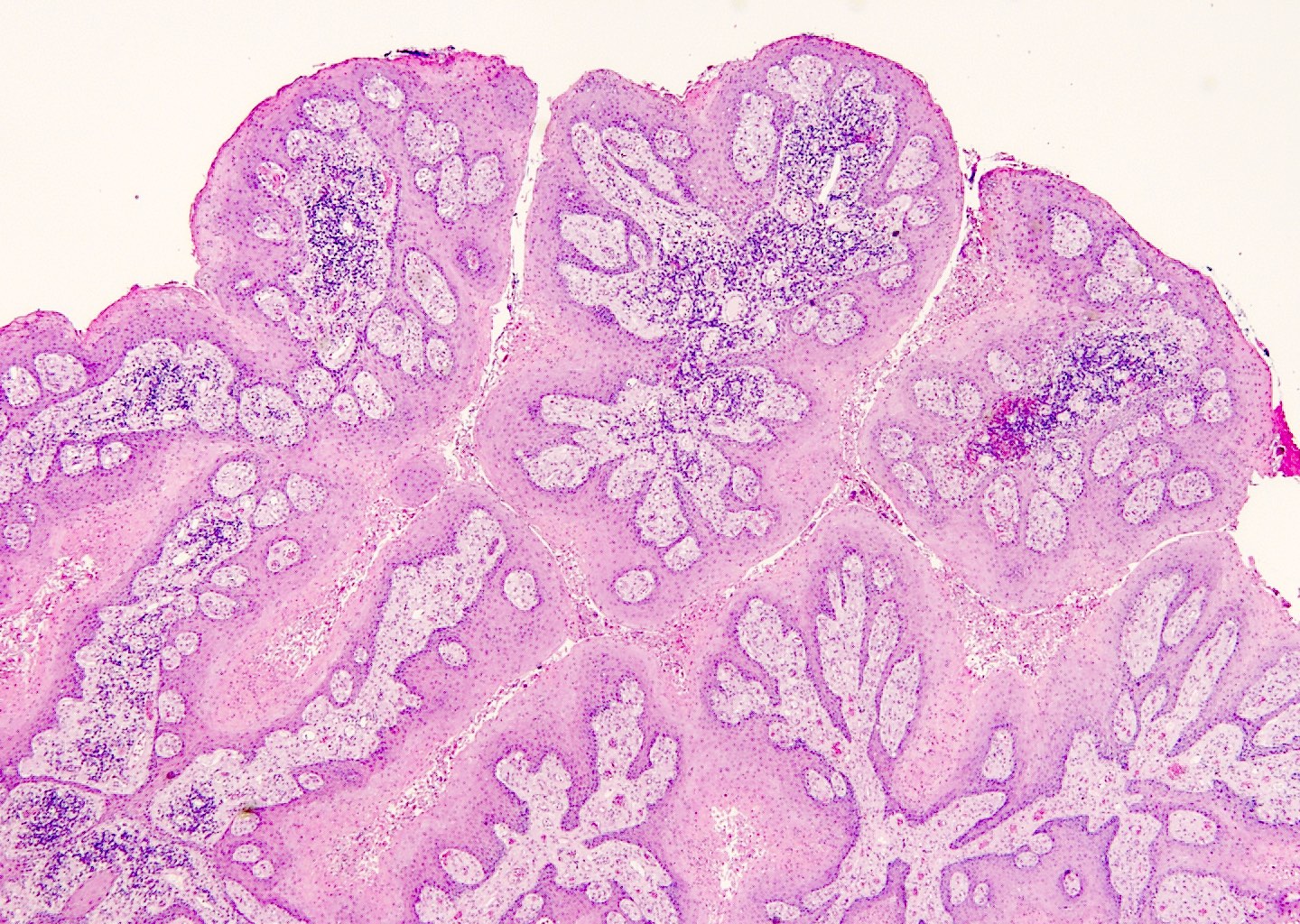
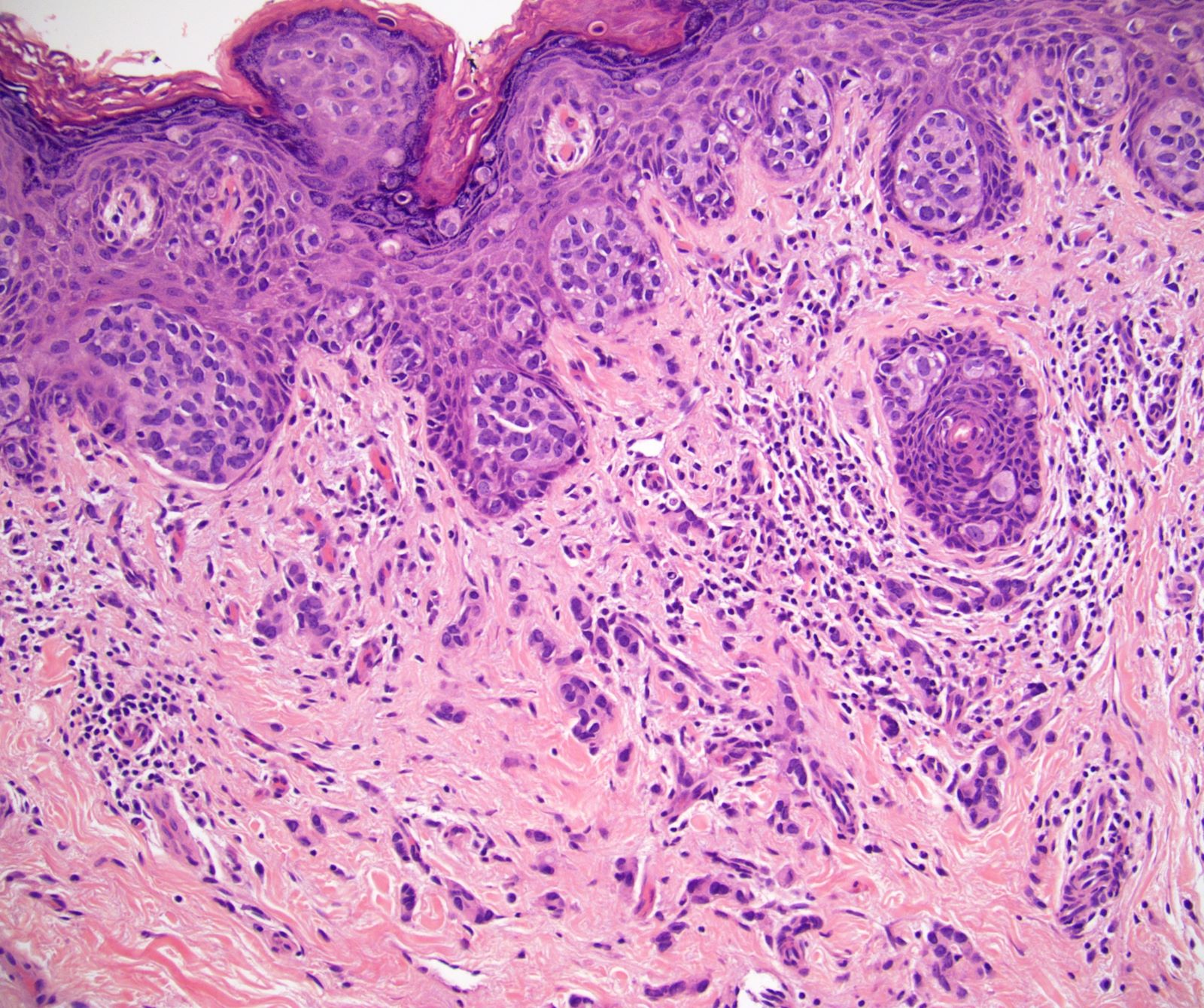
A 61 year old man with no cancer history had a flaky rash in the penoscrotal region for 9 months. CT scans of the abdomen and pelvis were negative. He underwent a scrotal excision and partial penectomy.

All cases are archived on our website. To view them sorted by case number, diagnosis or category, visit our main Case of the Month page.

To subscribe to Case of the Month or our other email newsletters, visit pathologyoutlines.com/subscribe.html.

2 June 2021: Pathology Images of the Week

Here is a selection of new images from our free, online textbook:

Lung > Lepidic adenocarcinoma

Noninvasive lepidic adenocarcinoma (left) juxtaposed with adjacent uninvolved lung parenchyma (right). The lepidic adenocarcinoma is characterized by thickened alveolar septa lined by atypical overlapping cuboidal cells.

Contributed by Jonathan Keow, M.D., Ph.D.

Skin nonmelanocytic tumor > Trichilemmal (pilar) type cysts

High power view of epithelial lining with maturation of squamous epithelium, which lacks a granular layer (trichilemmal differentiation).

Contributed by Aaron Muhlbauer, M.D. and Jodi Speiser, M.D.

Skin nontumor > Granuloma annulare

Palisaded granuloma annulare. Arm of a 50 year old woman.

Contributed by Stephen Somach, M.D.

Vulva, vagina & female urethra > Smooth muscle tumors

Leiomyosarcoma of the vulva, morphologically low grade, with moderately atypical, round to cigar shaped nuclei with focally prominent nucleoli (400x). Mitoses are rare (1 – 2 per 10 high power fields) and necrosis is absent.

Contributed by David B. Chapel, M.D.

Penis & scrotum > Cutaneous verruciform xanthoma

A low power view shows papillary / verrucous epithelial hyperplasia with band-like plasma cell infiltrate in the scrotum.

Contributed by Liwei Jia, M.D., Ph.D.

Autopsy & forensics > Blunt force injuries

Anterior cranial base fractures (yellow arrows).

Contributed by Lorenzo Gitto, M.D., Robert Stoppacher, M.D., Serenella Serinelli M.D., Ponni Arunkumar, M.D. and Luigi Bonaccorso, M.D.