
This was a 30 year old primi gravida with no history of consanguinity . This was referred for evaluation of suspected ventriculomegaly.
The findings were :
prominent ventriculomegaly of posterior horns of both lateral ventricles – colpocephaly
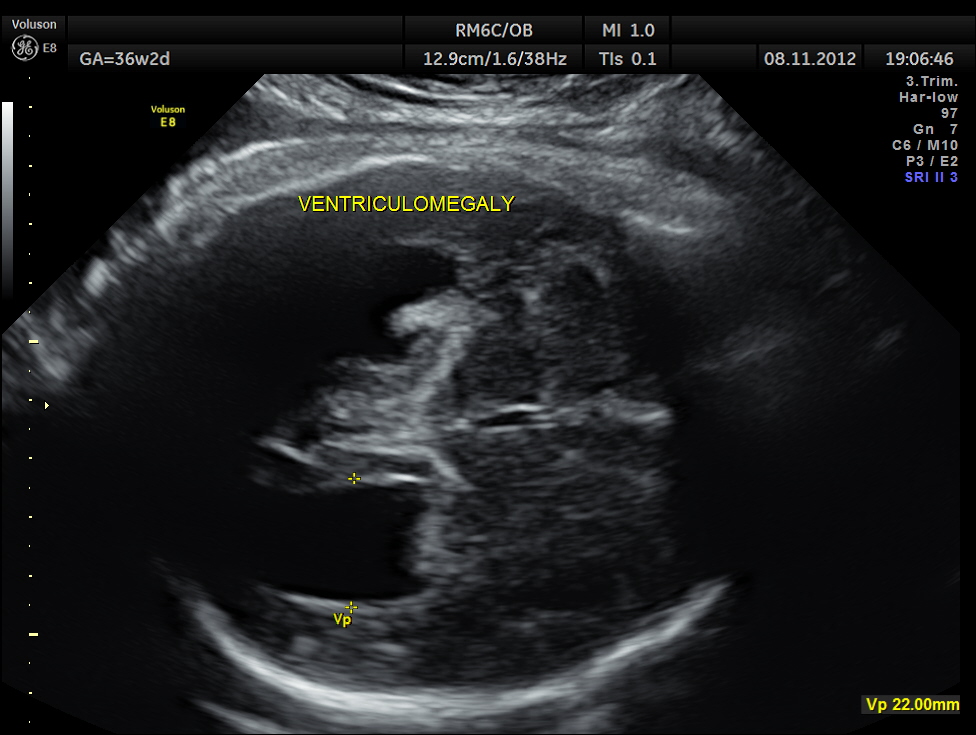
dilated occipital horns -colpocephaly

cavum septum pellucidum is not made out clearly

demonstration of both medial and lateral ventricular walls at a level where the single periventricular line is normally demonstrated


The following links would be very helpful to understand more :
http://www.sonoworld.com/fetus/page.aspx?id=107
http://www.fetalultrasound.com/online/text/5-077.HTM
A specific diagnosis of agenesis of the corpus callosum has seldom been made before the third trimester, probably because the corpus callosum is not normally formed until 18 to 20 weeks43. Most authors agree that detection of agenesis of the corpus callosum is difficult prenatally, depending as it does on postnatal sonograms or CT or MR scans. In a series of seven fetuses with agenesis of the corpus callosum, Bertino et al35 reported that only three demonstrated a characteristic midline cyst. They described three findings that might lead one to suspect agenesis of the corpus callosum on routine transverse views:
- disproportionate enlargement of the occipital horn,
- demonstration of both medial and lateral ventricular walls at a level where the single periventricular line is normally demonstrated, and
- a more parallel course of both ventricular walls than normal.
They suggested that demonstration of these findings on axial views should stimulate additional coronal and sagittal views for evaluation of agenesis of the corpus callosum.
The increased separation of the normal-sized bodies and the enlargement of the atria and occipital horns of the lateral ventricle result in a typical ultrasound image. Upward displacement of the third ventricle is a very specific sign36 but presents only in 40% of fetuses.

Also, on every Ob study, I was taught to make sure the CSP looked like a clear black box without any lines. Our radiologists wouldn’t pass the picture without that criteria being met.
LikeLike
Agreed
LikeLike
It seems that one must know what the lines represent before saying they should not be there. It is unclear what the motivation might be for a radiologist to refuse any picture without “lines” that may be in the picture. The mid cave position is one of several variants and can be demonstrated in more than one way.
LikeLike
Dear Sir, It’s a great work for reference for those who just entered in ULTRASOUND field….Excellent work….Sir……
LikeLike
I’m amazed, I have to admit. Seldom do I come
across a blog that’s both equally educative and engaging, and without a doubt, you have hit the
nail on the head. The issue is something that not enough men and women are speaking intelligently about.
I am very happy I came across this in my hunt for
something regarding this.
LikeLike
Thanks
LikeLike