uterus
(redirected from uterus duplex)Also found in: Dictionary, Thesaurus, Medical.
uterus
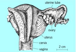
uterus, in most female mammals, hollow muscular organ in which the fetus develops and from which it is delivered at the end of pregnancy. The human uterus is pear-shaped and about 3 in. (7.6 cm) long (it expands greatly during pregnancy); it normally lies in the pelvis, where it is supported by a ligament on either side extending to the pelvic wall. The body of the uterus tapers down to a necklike structure (cervix) that leads into the vagina. On either side of the uterus is an oviduct (called fallopian tube, or uterine tube, in humans) from 3 to 5 in. (7.6–12.7 cm) long, one end opening into the uterus and the other, wide-mouthed, ends in close proximity to an ovary. These oviducts serve as passageways for the ova to reach the uterus. Fertilization occurs in the oviduct; the fertilized ovum then continues into the uterus, where it becomes implanted in the lining of that organ, also known as the endometrium. If fertilization does not occur, the ovum and the lining of the uterine wall pass out of the body through the vagina (see menstruation). Endometrial tissues then build up again in the uterus in anticipation of the next release of an ovum.
Diseases that can affect the uterus include various sexually transmitted diseases, pelvic inflammatory disease, and endometriosis, as well as benign or malignant tumors. Benign tumors may be removed without damage to the uterus, although in cases where the tissue is found to be cancerous, the entire uterus and cervix may have to be removed in a procedure known as hysterectomy. Prolapse of the uterus occurs when weakened supporting structures allow the uterus to tilt and slip downward into the vagina. See also reproductive system.
Uterus
The hollow, muscular womb, being an enlarged portion of the oviduct in the adult female. An adult human uterus, before pregnancy, measures 3 × 2 × 1 in. (7.5 × 5 × 2.5 cm) in size and has the shape of an inverted, flattened pear. The paired Fallopian tubes enter the uterus at its upper corners; the lower, narrowed portion, the cervix, projects into the vagina (see illustration). Normally the uterus is tilted slightly forward and lies behind the urinary bladder.
The lining, or mucosa, responds to hormonal stimulation, growing in thickness with a tremendous increase in blood vessels during the first part of the menstrual cycle. If fertilization does not occur, the thickened vascular lining is sloughed off, producing the menstrual flow at the end of the cycle, and a new menstrual cycle begins with growth of the mucosa. When pregnancy occurs, the mucosa continues to thicken and forms an intimate connection with the implanted and enlarging placenta. See Menstruation, Reproductive system
Uterus
in females a special, expanded section of the oviducts with a powerful muscular wall and a good supply of blood.
In animals. A uterus is found in annelids, arthropods, mollusks, most lower vertebrates (some cartilaginous and all viviparous bony fishes), certain amphibians, many reptiles, birds, and all mammals.
In oviparous vertebrates (reptiles and birds), the mature ova are held temporarily in the uterus. In viviparous animals, the embryonic development of the organism takes place there, using either the nutrients of the ovum or those of the mother. In the latter case, communication and the exchange of matter between the developing embryo and the uterus are accomplished through the placenta.
The structure of the uterus differs greatly from animal to animal. In Prototheria (duck-billed platypus, spiny anteater), the uterus is paired, and each uterus opens separately into a cloaca. In marsupials (kangaroo), the uterus is also paired, but each opens into a special vagina. In placental mammals, one finds all forms from paired uterus to unpaired.
Four main types of uterus are distinguished, depending on the degree of concrescence of the oviducts: uterus duplex, or two uteri, each opening independently into a common vagina (found in some rodents, elephants); uterus bipartitus, or two uteri, with the posterior sections grown together and opening by a common orifice into the vagina (in some rodents, ruminants, swine, carnivores); uterus bicornis, the most common in mammals, consisting of two uterine horns that join in the unpaired body of the uterus and opening into a vagina (in many carnivores, insectivores, cetaceans, artiodactyls, perissodactyls); and uterus simplex, consisting only of an unpaired body into which open two oviducts (in most chiropterans, primates, man).
REFERENCES
Kholodkovskii, N. A. Uchebnik zoologii, 6th ed. Moscow-Leningrad, 1933.Kurs zoologii, 7th ed., vols. 1-2. Moscow, 1966.
Marshall’s Physiology of Reproduction, 3rd ed., 2 vols. Edited by A. S. Parkes. London-New York-Toronto, 1956-58.
Giersberg, H., and P. Rietschel. Vergleichende Anatomie der Wirbeltiere, vol. 2. Jena, 1968.
K. M. KURNOSOV
In man. In the female, the uterus is the fetus-bearing hollow muscular organ in the lesser pelvic cavity between the urinary bladder and the rectum.
The human uterus weighs 40-50 g in nulliparas and 90-100 g in multiparas. The organ is pear-shaped; the larger part is made up of the body (the upper part of which, called the fundus, is enlarged). The lower, narrowed end of the uterus (cervix) is held by the vagina. The cervix consists of two parts: the vaginal (facing the vagina) and the supravaginal. The fundus of the uterus inclines forward; the body and the cervix form an angle that opens forward. The cavity inside the uterus is triangular, with two upward openings leading to the fallopian tubes. The cavity of the uterus passes the cervical canal, which opens at the uterine ostium into the vagina.
The wall of the uterus consists of three membranes: outer (serous), middle (muscular), and inner (mucous). The serous membrane is represented by the peritoneum, which envelops the uterus from the front, rear, and sides and passes to the urinary bladder and the rectum, circumscribing the vesicouterine and rectouterine pouches. Along the sides of the uterus, the sheets of the peritoneum grow together to form the broad ligament of the uterus, which, together with the fascia and muscles of the pelvic floor, help to secure the organ.
The middle layer of the uterus is the most powerful, consisting of three layers of smooth muscle with added elastic fibers. The mucous membrane is lined with ciliated cylindrical epithelium and holds numerous glands. This layer undergoes changes in connection with the menstrual cycle.
Arterial blood is supplied to the uterus by branches of the uterine and ovarian arteries, which are especially well developed during pregnancy. Venous blood leaves the uterus through the uterine and ovarian veins; lymph, through efferent vessels to the aortoabdominal, hypogastric, and iliac lymph nodes. The uterus
| Table 1. Output of principal products of the vegetable oil and fat industry in the USSR (thousands of tons) | |||||||
|---|---|---|---|---|---|---|---|
| 1913 | 1928 | 1940 | 1950 | 1960 | 1970 | 1972 | |
| Vegetable oil | 538 | 448 | 798 | 819 | 1,586 | 2,784 | 2,842 |
| Margarine | – | – | 121 | 192 | 431 | 762 | 850 |
| Mayonnaise | – | – | – | – | 7.8 | 40.8 | 52.8 |
| Soap (figured at 40-percent fatty-acid content) | 192 | 311 | 700 | 816 | 1,451 | 1,442 | 1,223 |
| Synthetic detergents | – | – | – | – | 22.9 | 470 | 534 |
| Natural drying oil | – | – | 36.6 | 15 | 63.1 | 44.9 | 41.4 |
| produced at enterprises of the Ministry of the Food Industry | 15.6 | 16.8 | |||||
is innervated by branches of the inferior mesenteric plexus and the pelvic nerves.
IA. L. KARAGANOV
