History
Because only approximately 25-30% of United States patients with early Lyme disease recall the tick bite, the clinician must direct the history toward the possibility of a tick bite. (In Europe, 64% do not remember being bitten.) Patients are generally unaware of a tick bite because these ticks are extremely small (nymphal Ixodes ticks are approximately the size of a poppy seed) and their bites are often painless.
Epidemiologic context is extremely important. The clinician should determine where the patient lives, works, and vacations, and should ask about specific activities in which the patient participates at those locales. The probability of a tick bite—and thus, the likelihood of contracting Lyme disease—is highest in persons who spend time outdoors (particularly in wooded, brushy, or grassy habitats) in a geographically endemic area.
Endemic areas can be defined as those with established populations of Ixodes scapularis or other vector ticks and evidence of enzootic transmission of Borrelia burgdorferi between the tick and the resident animal population. (See Epidemiology.)
The season is important, especially in patients with early disease. Most cases of erythema migrans occur from late spring through early fall, because that is when ticks in the nymphal stage are seeking a blood meal, and nymphs account for 90% of Lyme disease cases. For patients presenting with later cutaneous manifestations, especially acrodermatitis chronica atrophicans, questions must be directed at assessing the risk of tick bite (or previous manifestations of Lyme disease) from many years in the past.
Because people who engage in activities that put them in risk for tick bites tend to continue those activities, reinfection is not uncommon. Patients who have previously had erythema migrans can be reinfected (meaning that the first infection has been successfully treated, and they have a new infection with B burgdorferi). This has been clearly demonstrated in reinfected patients who were culture positive and had different serotypes isolated in the first and second infections.
Reinfections manifest in much the same ways as first infections, although a tendency towards less hematogenous spread is noted. In contrast, relapse (as opposed to reinfection) is very unusual in patients who have been treated with appropriate antimicrobials. [10]
Certain manifestations of Lyme disease are related to the particular strain of Borrelia involved. In the United States, isolates from the East Coast are known as B burgdorferi sensu stricto, and infection with this organism has a particular predilection to affect joints, whereas other strains are more common in Europe, such as B burgdorferi afzelii, which is associated with acrodermatitis chronica atrophicans.
Similar to syphilis, the manifestations of Lyme disease have been divided into three stages: localized, disseminated, and persistent. However, in individual patients, no rigid cutoffs exist between stages. The first two stages are part of the early infection, whereas persistent disease is considered late infection. Unlike syphilis, stage 3 disease may occur within 1 year of infection, not many years later.
Stage 1 Lyme disease
Early localized Lyme disease refers to isolated erythema migrans and to an undifferentiated febrile illness. This stage occurs 1-30 days after the tick bite.
Erythema migrans, the characteristic skin rash of Lyme disease, occurs in two thirds of patients with Lyme disease and develops at an average of 7 days after the tick bite. [5] The rash typically occurs at or near the site of the tick bite, which may be an area not normally visualized by individuals, such as the axilla, groin, or popliteal areas. It may be asymptomatic or it may itch or burn.
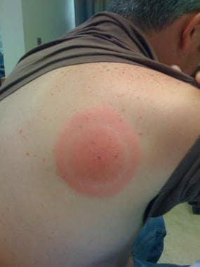
The rash typically expands over days and is not evanescent. It may not be observed until it is already full size. Clearing of portions of the rash as it expands may result in concentric rings of erythema, producing the classic bull’s-eye rash (see the image below). In the United States, however, erythema migrans is more likely to have a uniform color.
Although many patients present with erythema migrans, others first present with extracutaneous symptoms. In those cases, erythema migrans may have never occurred or may not have been recognized by the patient or correctly diagnosed by the physician.
Untreated, the rash may persist for 2-3 weeks. Eighty percent of patients with Lyme disease have only one episode of erythema migrans, whereas 20% may have recurrent episodes. Multiple lesions may occur in 20% of patients with Lyme disease; they result from hematogenous dissemination and are not the result of multiple tick bites (see the following image).
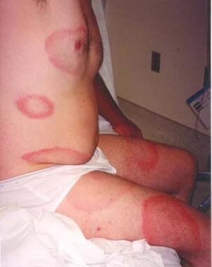 Lyme disease. Multiple lesions of erythema migrans occur in approximately 20% of patients. A carpenter from Nantucket who worked predominantly outside had been treated with clotrimazole/betamethasone for 1 week for a presumed tineal infection, but the initial lesion grew, and new ones developed. He then presented to the emergency department with the rashes seen in this photo. The patient had no fever and only mild systemic symptoms. He was treated with a 3-week course of oral antibiotics.
Approximately 50% of patients describe flulike symptoms within days to 1 week of infection, characterized by fever, chills, and malaise. Fever is generally low grade. Other symptoms include fatigue and myalgia (80% of patients with Lyme disease in the US but < 35% in Europe), as well as arthralgia, headache, and neck stiffness, which may resolve spontaneously even if specific therapy is not initiated. A paucity of respiratory and gastrointestinal tract symptoms may also be present. [33]
The most common ocular manifestations of stage 1 Lyme disease are redness and tearing.
Approximately one third of all patients with erythema migrans develop no further manifestations of Lyme disease. Two thirds of patients develop further symptoms (stages 2 and 3).
It is also important to consider co-infection by other organisms transmitted by the same tick bite, in areas where those are endemic. Co-infection with Ehrlichia species and Babesia microti are reported with increased frequency; in some studies, co-infection occurs in as many as 10-15% of patients with Lyme disease. When Lyme disease is strongly suggested but some of the manifestations are atypical (eg, a high fever, especially if accompanied by rigors or a toxic appearance) these other tick-borne infections or an alternative diagnosis must be considered.
Stage 2 Lyme disease
Early disseminated Lyme disease usually develops 3-10 weeks after inoculation. Approximately 25% of individuals infected with B burgdorferi have signs and symptoms of disseminated disease at presentation.
Systemic manifestations may include fever and malaise. One or more organ systems become involved as hematologic or lymphatic spread disseminates spirochetes to distant sites. Musculoskeletal and neurologic symptoms are the most common; less common are symptoms from cardiac disturbances, such as dizziness, syncope, dyspnea, chest pain, and palpitations.
Ocular manifestations include diplopia secondary to a cranial neuropathy or Bell palsy. Blurred vision and eye pain can occur from keratitis and iritis. Unilateral blindness from panophthalmitis has been reported as well.
Musculoskeletal manifestations
Intermittent inflammatory arthritis often begins as a migratory polyarticular process involving bursae, tendons, and joints, which evolves over 1-2 days into a monoarticular process involving the knee, ankle, and wrist, in decreasing frequency. When asked about symptoms after they have resolved, patients with Lyme disease are less likely to remember those symptoms that occurred before the monoarthritis. Polyarticular episodes may also occur.
In two thirds of patients, the first episode occurs within 6 months of the erythema migrans lesion. Untreated, the episodes last approximately 1 week. Two thirds of patients have three recurrences approximately 2.5 months apart. The recurrences are more likely to involve more than one joint. With time, these episodes become less frequent and severe and involve fewer joints. Even without treatment, the recurrent episodes usually resolve over a 10-year period.
Some patients may present with intermittent joint pain without inflammatory findings. This is more common in Europe, where arthritis was not recognized as a manifestation of Lyme disease until the early reports from the United States.
Neurologic manifestations
Neurologic involvement, also known as Lyme neuroborreliosis, is reported in 5-20% of cases. In the United States, cranial neuropathy is the most common manifestation of early neurologic Lyme disease. Other manifestations include meningitis and encephalopathy.
Cranial neuropathies, especially facial nerve palsy (Bell palsy), develop in approximately 3% of Lyme disease patients. In endemic areas, Lyme disease is the most commonly identified cause of acquired facial palsy, especially in children. [34] Headache, absence of previous herpetic lesions, and meningeal symptoms are noted in most pediatric Lyme disease patients with facial palsy. [34]
When meningitis is involved, symptoms usually occur 2-10 weeks following infection. Headache, neck pain or stiffness, and photophobia typically indicate meningeal irritation. The headache of Lyme disease typically is described as waxing and waning, and the severity varies from mild to severe, even in patients with frank meningitis. Persistent headache alone is a rare presentation of Lyme disease but should be considered in patients in endemic areas during summertime.
Borrelia encephalopathy most commonly manifests as a mild confusional state accompanied by disturbances in memory, concentration, mood, sleep, personality, and/or language occurring months to years after the infection. Depression and irritability are also common.
Cutaneous manifestations
In patients with cutaneous involvement, multiple erythema migrans lesions are present. These are relatively small erythematous macules (1-5 cm) and are often oval. Unlike primary single erythema migrans rashes, these lesions can be evanescent and do not show the typical expansion over days.
Borrelial lymphocytoma is an uncommon manifestation of early disseminated Lyme disease (though it may also occur very early in the disease course) that has been reported only from Europe. It is a bluish-red nodular swelling that typically occurs on the lobe of the ear in children (see the following image) or the areola of the nipple in adults. Occasionally, borrelial lymphocytoma lesions occur on the scrotum, nose, and extremities. Nipple lesions tend to be painful, possibly because of rubbing against clothing.
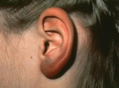 Lyme disease. Borrelial lymphocytoma of the earlobe, which shows a bluish red discoloration. The location is typical in children, as opposed to the nipple in adults. This manifestation of Lyme disease is uncommon and occurs only in Europe. Courtesy of Lyme Disease Foundation, Hartford, Conn.
Stage 3 Lyme disease
Late or chronic Lyme disease refers to manifestations that occur months to years after the initial infection, sometimes after a period of latency. Signs and symptoms of chronic Lyme disease are primarily rheumatologic and neurologic. Acrodermatitis chronica atrophicans, the cutaneous feature of late-stage Lyme disease, is found almost exclusively in European patients.
Most patients presenting with late disease do not have a history of erythema migrans, because the rash typically leads to earlier treatment, which prevents the development of late disease. However, other manifestations of the disease may coexist or may have occurred in the past. Thus, a history of Bell palsy, aseptic meningitis, arthritis, acral paresthesias or dysesthesias (from peripheral neuropathy), or cognitive dysfunction (from CNS involvement) may be diagnostically useful.
Maladaptive host responses can lead to a variety of syndromes in stage 3, as follows [35] :
-
Postinfectious Lyme arthritis – Massive inflammatory synovial proliferation, usually in a knee
-
Posttreatment Lyme disease syndrome – Pain, neurocognitive impairment, fatigue
-
Autoimmune joint disease – Rheumatoid arthritis, psoriatic arthritis, or peripheral spondyloarthropathy
-
Autoimmune neurologic disease – Chronic idiopathic demyelinating polyneuropathy
Lyme arthritis is the hallmark of stage 3 Lyme disease. It tends to involve large joints (the knee is involved in 90% of cases). Arthritis must be differentiated from arthralgia, which is common in early disease.
The neurologic abnormalities of stage 3 Lyme disease involve both the central and peripheral nervous systems. Typical presentations include subacute encephalopathy, chronic progressive encephalomyelitis, and late axonal neuropathies, as well as symptoms consistent with fibromyalgia. Radicular pain can occur and present as acute disk disease. More common in European patients, radicular pain may be associated with lymphocytic pleocytosis (Bannwarth syndrome).
Borrelia encephalomyelitis is a rare but severe syndrome. Symptoms can progress gradually or in a relapsing-remitting pattern, with partial improvement after the attacks. The most common clinical manifestations of Borrelia encephalomyelitis are hemiparesis, ataxia, seizures, cognitive impairment, bladder dysfunction, and hearing loss. Myelitis is present in 50% of patients with late neuroborreliosis. Progressive spastic paraparesis or quadriparesis is common.
Acrodermatitis chronica atrophicans most commonly affects older women. Unlike erythema migrans, acrodermatitis chronica atrophicans tends to occur acrally, especially on the dorsal surfaces of the hands, feet, knees, and elbows. Early on, a minimally symptomatic erythema tends to occur in these locations. Initially, there is discoloration and inflammation; later, severe atrophy is noted. [15] See the image below.
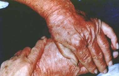 Acrodermatitis chronica atrophicans is found almost exclusively in European patients and comprises an early inflammatory phase and a later atrophic phase. As the term suggests, the lesion occurs acrally and ultimately results in skin described as being like cigarette paper. Courtesy of Lyme Disease Foundation, Hartford, Conn.
Physical Examination
In many patients with early Lyme disease, identification of erythema migrans (EM) on physical examination alone is sufficient to establish a working diagnosis of Lyme disease. Careful attention to the details often makes the difference between the need to proceed with further confirmatory tests and an empiric course of antibiotics. In particular, the examination findings must be interpreted in the epidemiologic context; this cannot be overemphasized. The location, time of year, and patient's activities can be important diagnostic clues.
Regional lymphadenopathy may be seen, and a low-grade fever is not uncommon. High fever suggests another co-infecting tick-borne organism such as Ehrlichia or Babesia species or some other diagnosis altogether, such as streptococcal cellulitis.
Erythema migrans
EM is the characteristic rash of Lyme disease. Classic EM is a flat to slightly raised erythematous lesion that appears at the site of the tick bite after 1-33 days bite (average, 7-10 days). [36] Without therapy, erythema migrans typically fades within 3-4 weeks.
EM usually is round or oval, but can be triangular or linear. Often, a central punctum is evident at the bite site.
EM enlarges by a few centimeters per day; single lesions typically achieve a diameter of approximately 16 cm (approximately 5-6 inches), but lesions as large as 70 cm have been reported. The case definition for Lyme disease used by the US Centers for Disease Control and Prevention (CDC) specifies that EM be greater than 5 cm in size. This size cutoff is only meant to be used for epidemiologic purposes, however; EM smaller than 5 cm has occasionally been documented in culture-proven cases. [37]
The entire lesion may be uniform in color or have central darkening. Central clearing is more common in European patients than in North American patients. More proximal to the clearing may be additional erythema leading to a so-called bull's eye or target appearance; however, this phenomenon, emphasized in the earlier literature, occurs only in a minority of patients (37% in one North American study of culture-proven EM). [38] See the images below.
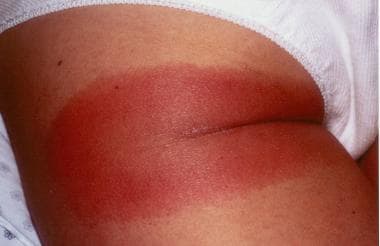 Lyme disease. This patient's erythema migrans rash demonstrates several key features of the rash, including size, location, and presence of a central punctum, which can be seen right at the lateral margin of the inferior gluteal fold. Note that the color is uniform; this pattern probably is more common than the classic pattern of central clearing. On history, this patient was found to live in an endemic area for ticks and to pull ticks off her dog daily.
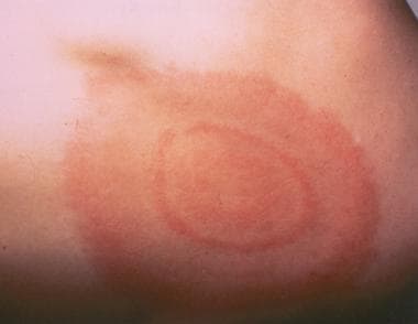 Lyme disease. Classic target lesion with concentric rings of erythema, which often show central clearing. Although this morphology was emphasized in earlier North American literature, it only represents approximately 40% of erythema migrans lesions in the United States. This pattern is more common in Europe. Courtesy of Lyme Disease Foundation, Hartford, Conn.
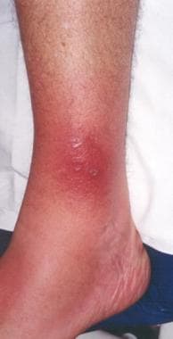 Lyme disease. The rash on the ankle seen in this photo is consistent with both cellulitis (deep red hue, acral location, mild tenderness) and erythema migrans (presentation in July, in an area highly endemic for Lyme disease). In this situation, treatment with a drug that covers both diseases (eg, cefuroxime or amoxicillin-clavulanate) is an effective strategy.
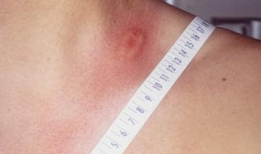
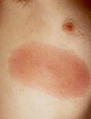 Lyme disease. The thorax and torso are typical locations for erythema migrans. The lesion is slightly darker in the center, a common variation. In addition, this patient worked outdoors in a highly endemic area. Physical examination also revealed a right axillary lymph node.
Atypical manifestations of EM include vesicular (see the image below) and centrally necrotic lesions. Scaling is unusual, but may be seen, especially in the center of the lesion.
 Lyme disease. Photo of the left side of the neck of a patient who had pulled a tick from this region 7 days previously. Note the raised vesicular center, which is a variant of erythema migrans. The patient had a Jarisch-Herxheimer reaction approximately 18 hours after the first dose of doxycycline.
Rash location is another important diagnostic clue. Unlike spider and other arthropod bites, EM rarely is found on the hands or feet. Rather, ticks tend to bite where natural barriers impede their forward motion (eg, popliteal fossa, axillary or gluteal folds, hairline, areas near elastic bands in bra straps or underwear). In children, the scalp, face, and hairline are especially common locations. [8, 39]
Approximately 20% of patients with EM have secondary lesions (see the image below). These lesions generally are smaller than the primary one, lack the central punctum, and are not necrotic or vesicular. They also tend to be more uniform in morphology than the primary lesion. Because secondary lesions result from hematogenous spread, their locations are not as restricted as those of the primary lesion.
Lyme disease. Multiple lesions of erythema migrans occur in approximately 20% of patients. A carpenter from Nantucket who worked predominantly outside had been treated with clotrimazole/betamethasone for 1 week for a presumed tineal infection, but the initial lesion grew, and new ones developed. He then presented to the emergency department with the rashes seen in this photo. The patient had no fever and only mild systemic symptoms. He was treated with a 3-week course of oral antibiotics.
Note that rashes very similar to erythema migrans, but from which Borrelia burgdorferi cannot be cultured, have been reported in the southern United States. This disease is called southern tick-associated rash illness (STARI), or Master disease. As a group, distinctions can be made between classic erythema migrans and this illness, but significant overlaps exist such that the differences are not useful in diagnosing individual patients.
An erythematous skin lesion present while an Ixodes tick is still attached is most likely a hypersensitivity reaction rather than EM. Hypersensitivity reactions also tend to produce lesions that are smaller and more transient than EM; typically, the lesions are less than 5 cm in size and begin to resolve within 2 days.
Borrelial lymphocytoma
Less than 1% of patients with stage 2 Lyme disease, almost all of them European, develop Borrelia lymphocytoma, described as a small, bluish-red nodule or plaque. The earlobe and scrotum are the typical location in children (See the image below), whereas the nipple is the more common location in adults.
Lyme disease. Borrelial lymphocytoma of the earlobe, which shows a bluish red discoloration. The location is typical in children, as opposed to the nipple in adults. This manifestation of Lyme disease is uncommon and occurs only in Europe. Courtesy of Lyme Disease Foundation, Hartford, Conn.
Borrelial lymphocytoma tends to occur in areas of previous (or concurrent) erythema migrans and may be up to a few centimeters in size. Regional lymphadenopathy may be present.
Other terms used to describe borrelial lymphocytoma include the following:
-
Lymphadenosis benigna cutis
-
Lymphocytoma cutis
-
Cutaneous lymphoid hyperplasia
-
Spiegler-Fendt lymphoid hyperplasia
Acrodermatitis chronica atrophicans
Acrodermatitis chronica atrophicans is a relatively uncommon physical manifestation of late Lyme disease that begins as an inflammatory phase marked by edema and erythema, usually on the distal extremities; at times, a faint bluish discoloration is found predominantly on extensor surfaces. The lesions have a predilection for the posterior heels and dorsal (extensor) surfaces of the hands, feet, elbows, and knees. The lesions also tend to become symmetric. The buttocks often become involved.
Later, atrophy supervenes and so-called cigarette-paper skin is seen, with an appearance similar to scleroderma. (See the image below.) Because of the loss of subcutaneous fat, underlying venous structures are more visible, and the skin becomes thin, atrophic, and dry. Fibrous juxtaarticular nodules or bands may be seen on the extensor surfaces of the elbows and knees. One third of patients with acrodermatitis chronicum have an associated sensory polyneuropathy.
Acrodermatitis chronica atrophicans is found almost exclusively in European patients and comprises an early inflammatory phase and a later atrophic phase. As the term suggests, the lesion occurs acrally and ultimately results in skin described as being like cigarette paper. Courtesy of Lyme Disease Foundation, Hartford, Conn.
Other dermatologic manifestations
Other skin lesions have been associated with B burgdorferi infection, but whether they are part of the syndrome of Lyme disease is controversial. The lesion for which the most evidence of causality has been reported is morphea (localized scleroderma), which develops in roughly 10% of European patients with borrelial lymphocytoma and acrodermatitis chronica atrophicans.
Other European reports less commonly link the following with B burgdorferi infection:
-
Progressive facial hemiatrophy (Parry-Romberg syndrome)
-
Eosinophilic fasciitis
Musculoskeletal findings
Muscle tenderness can result from myositis. Tenderness of tendons and periarticular structures may be present.
Frank arthritis can occur after weeks, months, or years and may lead to erythema, edema, synovial effusion, and tenderness of the affected joints. Usually, this is a monoarthritis or oligoarthritis involving large joints, especially the knee. Swelling often is disproportional to the tenderness.
Lyme arthritis
Approximately 60% of all untreated patients develop symptoms of intermittent migratory monoarthritis. Episodes last a mean of 3 months and very often affect the knee or temporomandibular joints, although not universally. Migratory oligoarthritis involving the small or large joints can also occur. Joint symptoms develop in approximately 80% of all untreated patients within 2 years of infection.
The severity of joint involvement can range from intermittent episodes of subjective pain to frank arthritis to chronic erosive synovitis. During the attacks, the joints are swollen, hot, and painful, but they are not usually red or as severe as in a septic joint. Effusions may be large and generally recur following aspiration, as is often seen in spondyloarthropathies. Fewer than 10% of patients with arthritic sequelae develop pannus or erosion of cartilage and bone.
Neurologic involvement
Approximately 5-10% of untreated patients with Lyme disease have signs of cranial neuropathies, and up to 60% of patients with early neuroborreliosis develop cranial neuritis. Seventh nerve palsy is by far the most common. Bilateral facial palsy can be seen in 35% of patients and is a unique characteristic that is useful for distinguishing it from idiopathic Bell palsy and other disorders.
Typical associated findings depend on the nerve affected and can include visual or auditory disturbances, facial paresthesia, and/or vertigo. Other neurologic manifestations include diffuse or focal mononeuropathy multiplex (multifocal involvement of anatomically unrelated nerves), plexopathy, and/or radiculoneuropathy (more common in Europe). Less common presentations include myositis, pseudotumor cerebri, and cerebellitis.
Lyme meningitis is relatively common, occurring in as many as 15% of untreated patients bitten by the Ixodes tick and in 30% of Lyme disease cases, and does not manifest as the usual signs of bacterial meningitis (eg, boardlike rigidity, Kernig and Brudzinski signs). Meningitis may be accompanied by cranial or peripheral radiculoneuropathy. Neck stiffness can occur early, with or without frank meningitis.
Acute radiculoneuritis is reported in 50-85% of cases. Acute onset of motor deficits, severe radicular pain, and sensory loss are commonly seen after 2-4 weeks of infection. Multifocal asymmetric weakness is a common presentation. Although the presentation of inflammatory radiculoneuropathy is often indistinguishable from that of spinal-root compression, involvement of multiple dermatomes in the thorax and a lack of a precipitating injury can aid in diagnosis.
Chronic radicular paresthesias are usually not associated with motor or sensory deficits. The physical examination results are normal.
With peripheral neuropathy, patients usually report intermittent paresthesias. The most frequent finding upon examination is decreased vibratory sensation of the distal lower extremities. A stocking-glove distribution of epicritic sensory deficits is also a common finding. Sensory findings are more pronounced than motor findings.
With late axonal neuropathy, patients can report intermittent distal limb paresthesias months to years after infection. It is distinct from the neuropathy of early Lyme disease, because the symptoms are less severe. Acrodermatitis chronica atrophicans–associated neuropathy is common in Europe and manifests as neuropathic pain, paresthesias, and muscle cramps.
In Europe, common manifestations of Garin-Boujadoux-Bannwarth syndrome (Bannwarth syndrome) include neuritic pain, cranial neuritis without headache, and lymphocytic pleocytosis. Bannwarth syndrome has also been called tick-borne meningopolyneuritis, lymphocytic meningoradiculitis, and chronic lymphocytic meningitis. While this manifestation is typically associated with infection with B garinii, a recent cluster was reported from the Mayo Clinic in Rochester, MN. [40]
Neuropsychiatric findings in late-stage disease may include the following:
-
Depression
-
Anxiety
-
Schizophrenia-like psychosis
-
Bipolar disorder
-
Dementia
Cardiac involvement
Cardiac involvement ranges from atrial or ventricular arrhythmias to transient heart block or myopericarditis. Approximately 8% of untreated patients have acute-onset atrioventricular conduction abnormalities. Most cardiac episodes are isolated and transient, lasting less than a week. In rare instances, patients with heart block require electrical pacing.
In patients with complete heart block, cannon A waves may be observed in the neck. A slow or irregular pulse may be palpated. A cardiac rub, S3 and/or S4, may be auscultated in patients with myocarditis or pericarditis. Signs of tamponade very rarely can occur. In patients with chronic cardiac involvement with heart failure, typical signs of chronic heart failure may be present.
Ophthalmic involvement
Ophthalmic manifestations vary by disease stage. In stage 1 Lyme disease, the ocular manifestations are conjunctivitis and photophobia. These are mild and transient, and ophthalmologists usually need not be consulted.
Significant ophthalmic complications may appear during stage 2 Lyme disease. [41, 42, 43, 44] Blurred vision can be noted during stage 2, secondary to papilledema, optic atrophy, optic or retrobulbar neuritis, or pseudotumor cerebri. Optic nerve disease may be unilateral or bilateral, and solitary or associated with other neurologic or neuro-ophthalmologic manifestations. Some evidence exists that children are more predisposed to optic nerve disease than adults. [45]
In late stage 2 or stage 3 Lyme disease, most of the severe ocular manifestations of the disease are seen. These include the following:
-
Episcleritis
-
Symblepharon
-
Keratitis
-
Iritis
-
Posterior or intermediate uveitis
-
Pars planitis
-
Vitreitis
-
Chorioretinitis
-
Exudative retinal detachment
-
Retinal pigment epithelial detachment
-
Cystoid macular edema
-
Branch artery occlusion
-
Retinal vasculitis
-
Orbital myositis
-
Cranial nerve palsies
Of this group, keratitis, vitreitis, and pars planitis are the most common. The keratitis usually is a bilateral, patchy, nummular stromal keratitis. Posterior segment inflammatory disease generally presents as a bilateral pars planitis associated with granulomatous iritis and vitreitis. Many of these patients also have granulomatous keratic precipitates and posterior synechiae. [46]
-
Lyme disease. The bacterium Borrelia burgdorferi (darkfield microscopy technique, 400X; courtesy of the US Centers for Disease Control and Prevention).
-
Lyme disease. Magnified ticks at various stages of development.
-
Ticks are the most common vectors for vector-borne diseases in the United States. In North America, tick bites can cause Lyme disease, human granulocytic and monocytic ehrlichiosis, babesiosis, relapsing fever, Rocky Mountain spotted fever, Colorado tick fever, tularemia, Q fever, and tick paralysis. Europe has a similar list of illnesses caused by ticks, but additional concerns include boutonneuse fever and tick-borne encephalitis. Lyme disease is one of the most prominent tick-borne diseases, and its main vector is the tick genus Ixodes, primarily Ixodes scapularis. Image courtesy of the US Centers of Disease Control and Prevention.
-
Lyme disease. Approximate US distribution of Ixodes scapularis. Image courtesy of the US Centers for Disease Control and Prevention.
-
Lyme disease. In general, Ixodes scapularis must be attached for at least 24 hours to transmit the spirochete to the host mammal. Prophylactic antibiotics are more likely to be helpful if feeding is longer. This photo shows 2 I scapularis nymphs. The one on the right is unfed; the other has been feeding for 48 hours. Note its larger size and the fact that the midgut diverticula (delicate brown linear areas on the body) are blurred. Photo by Darlyne Murawski; reproduced with permission.
-
Lyme disease. Normal and engorged Ixodes ticks.
-
Amblyomma americanum is the tick vector for monocytic ehrlichiosis and tularemia. An adult and a nymphal form are shown (common match shown for size comparison). Image by Darlyne Murawski; reproduced with permission.
-
Approximate US distribution of Amblyomma americanum. Image courtesy of the US Centers for Disease Control and Prevention.
-
The soft-bodied tick of the genus Ornithodoros transmits various Borrelia species that cause relapsing fever. Photo courtesy of Julie Rawlings, MPH, Texas Department of Health. Relapsing fever is characterized by recurrent acute episodes of fever (usually >39°C). It is a vector-borne illness spread by lice and ticks. The spirochete species Borrelia is responsible.
-
The Ixodes scapularis tick is considerably smaller than the Dermacentor tick. The former is the vector for Lyme disease, granulocytic ehrlichiosis, and babesiosis. The latter is the vector for Rocky Mountain spotted fever. This photo displays an adult I scapularis tick (on the right) next to an adult Dermacentor variabilis; both are next to a common match displayed for scale. Photo by Darlyne Murawski; reproduced with permission.
-
Approximate US distribution of Dermacentor andersoni. Image courtesy of the US Centers for Disease Control and Prevention.
-
Rhipicephalus ticks are vectors for babesiosis and rickettsial infections, among others. Image courtesy of Dirk M. Elston, MD. In typical practice, testing ticks for tick-borne infectious organisms is not generally recommended. However, healthcare practitioners should become familiar with the clinical manifestations of tick-borne diseases (eg, Lyme disease, especially those practicing in endemic areas) and maintain a high index of suspicion during warmer months. Ticks can be placed in a sealed container with alcohol if they need to be transported and identified.
-
To remove a tick, use fine-tipped forceps and wear gloves. Grasp the tick as close to the skin surface as possible, including the mouth parts, and pull upward with steady, even traction. Do not twist or jerk the tick because this may cause the mouth parts to break off and remain in the skin; however, note that the mouth parts themselves are not infectious. When removing, wear gloves to avoid possible infection.
-
Lyme disease. This patient's erythema migrans rash demonstrates several key features of the rash, including size, location, and presence of a central punctum, which can be seen right at the lateral margin of the inferior gluteal fold. Note that the color is uniform; this pattern probably is more common than the classic pattern of central clearing. On history, this patient was found to live in an endemic area for ticks and to pull ticks off her dog daily.
-
Erythema migrans, the characteristic rash of early Lyme disease.
-
Lyme disease. The thorax and torso are typical locations for erythema migrans. The lesion is slightly darker in the center, a common variation. In addition, this patient worked outdoors in a highly endemic area. Physical examination also revealed a right axillary lymph node.
-
Lyme disease. Photo of the left side of the neck of a patient who had pulled a tick from this region 7 days previously. Note the raised vesicular center, which is a variant of erythema migrans. The patient had a Jarisch-Herxheimer reaction approximately 18 hours after the first dose of doxycycline.
-
Lyme disease. Classic target lesion with concentric rings of erythema, which often show central clearing. Although this morphology was emphasized in earlier North American literature, it only represents approximately 40% of erythema migrans lesions in the United States. This pattern is more common in Europe. Courtesy of Lyme Disease Foundation, Hartford, Conn.
-
Typical appearance of erythema migrans, the bull's-eye rash of Lyme disease.
-
Lyme disease. Bulls-eye rash.
-
Lyme disease. Photo of erythema migrans on the right thigh of a toddler. The size and location are typical of erythema migrans, as is the history of the patient vacationing on Fire Island, NY, in the month of August. No tick bite had been noted at this location. Approximately 25% of patients with Lyme disease are children, which is the same percentage of patients who do not recall a tick bite. Courtesy of Dr John Hanrahan.
-
Lyme disease. Multiple lesions of erythema migrans occur in approximately 20% of patients. A carpenter from Nantucket who worked predominantly outside had been treated with clotrimazole/betamethasone for 1 week for a presumed tineal infection, but the initial lesion grew, and new ones developed. He then presented to the emergency department with the rashes seen in this photo. The patient had no fever and only mild systemic symptoms. He was treated with a 3-week course of oral antibiotics.
-
Lyme disease. The rash on the ankle seen in this photo is consistent with both cellulitis (deep red hue, acral location, mild tenderness) and erythema migrans (presentation in July, in an area highly endemic for Lyme disease). In this situation, treatment with a drug that covers both diseases (eg, cefuroxime or amoxicillin-clavulanate) is an effective strategy.
-
Lyme disease. Borrelial lymphocytoma of the earlobe, which shows a bluish red discoloration. The location is typical in children, as opposed to the nipple in adults. This manifestation of Lyme disease is uncommon and occurs only in Europe. Courtesy of Lyme Disease Foundation, Hartford, Conn.
-
A rarely reported noninfectious complication for tick bites is alopecia. It can begin within a week of tick removal and typically occurs in a 3- to 4-cm circle around a tick bite on the scalp. A moth-eaten alopecia of the scalp caused by bites of Dermacentor variabilis (the American dog tick) has also been described. No particular species appears more likely to cause alopecia. Hair regrowth typically occurs within 1-3 months, although permanent alopecia has been observed.
-
Acrodermatitis chronica atrophicans is found almost exclusively in European patients and comprises an early inflammatory phase and a later atrophic phase. As the term suggests, the lesion occurs acrally and ultimately results in skin described as being like cigarette paper. Courtesy of Lyme Disease Foundation, Hartford, Conn.
-
Blood smear showing likely babesiosis. Babesiosis can be difficult to distinguish from malaria on a blood smear.
-
Life cycle of the Ixodes dammini tick. Courtesy of Elsevier.
-
Lyme disease in the United States is concentrated heavily in the northeast and upper Midwest; it does not occur nationwide. Dots on the map indicate the infected person's county of residence, not the place where they were infected. Courtesy of the US Centers for Disease Control and Prevention (CDC).
Tables
- Table 1. Clinical presentation and therapy for the stages of Lyme disease
- Table 2. Adult and pediatric treatment options, dosages, and routes of administration
- Table 3. Comparison of Infectious Diseases Society of America (IDSA) and International Lyme and Associated Diseases Society (ILADS) recommendations for Lyme disease treatment
Clinical Manifestations |
Adult Dose |
Pediatric Dose |
Erythema migrans |
Doxycycline 100 mg PO BID for 10-14 days, OR
Amoxicillin 500 mg PO TID for 14 days, OR
Cefuroxime axetil 500 mg PO BID for 14 days
Patients unable to take doxycycline or beta-lactam antibiotics: Azithromycin 500 mg PO qDay for 7 days |
Doxycycline 4.4 mg/kg/day PO, divided into 2 doses; not to exceed 100 mg/dose for 10-14 days, OR
Amoxicillin 50 mg/kg/day PO, divided into 3 doses; not to exceed 500 mg/dose for 14 days, OR
Cefuroxime axetil 30 mg/kg/day PO, divided into 2 doses; not to exceed 500 mg/dose for 14 day |
Facial palsy [3] |
Doxycycline 100 mg PO BID for 14-21 days |
Doxycycline 4.4 mg/kg/day PO, divided into 2 doses; not to exceed 100 mg/dose for 14-21 days |
Lyme meningitis or radiculoneuritis [3] |
Doxycycline 200 mg/day PO, divided into 1-2 doses for 14-21 days, OR
Ceftriaxone 2 g IV qDay for 14-21 days; may substitute for oral therapy once the patient is stabilized or discharged to complete the course |
Doxycycline 4.4 mg/kg/day PO, divided into 1-2 doses; not to exceed 100 mg/dose for 14-21 days, OR
Ceftriaxone 50-75 mg/kg IV qDay; not to exceed 2 g/day for 14-21 days; may substitute for oral therapy once the patient is stabilized or discharged to complete the course |
Lyme disease–associated meningitis, cranial neuropathy, radiculoneuropathy, or with other peripheral nervous system (PNS) manifestations |
Without parenchymal involvement of brain or spinal cord: IV ceftriaxone, cefotaxime, penicillin G, or oral doxycycline With parenchymal involvement of brain or spinal cord: IV antibiotics are preferred |
|
Mild lyme carditis (1st degree AV block with PR interval < 300 milliseconds) [3] |
Doxycycline 100 mg PO BID for 14-21 days, OR
Amoxicillin 500 mg PO TID for 14-21 days, OR Cefuroxime axetil 500 mg PO BID for 14-21 days |
Doxycycline 4.4 mg/kg/day PO, divided into 2 doses; not to exceed 100 mg/dose for 14-21 days, OR
Amoxicillin 50 mg/kg/day PO, divided into 3 doses; not to exceed 500 mg/dose for 14-21 days, OR
Cefuroxime axetil 30 mg/kg/day PO, divided into 2 doses; not to exceed 500 mg/dose for 14-21 days |
Severe lyme carditis (symptomatic, 1st degree AV block with PR interval ≥300 milliseconds, 2nd or 3rd degree AV block) [3] |
Ceftriaxone 2 g IV qDay for 14-21 days; once symptoms and high-grade AV block resolve, consider transitioning to oral antibiotics to complete treatment course |
Ceftriaxone 50-75 mg IV qDay; not to exceed 2 g/day for 14-21 days; once symptoms and high-grade AV block resolve, consider transitioning to oral antibiotics to complete treatment course |
Borrelial lymphocytoma |
Oral antibiotic therapy for 14 days |
|
Arthritis |
Doxycycline 100 mg PO BID for 28 days, OR
Amoxicillin 500 mg PO TID for 28 days, OR
Cefuroxime axetil 500 mg PO BID for 28 days |
For ≥8 years:
Doxycycline 4.4 mg/kg/day PO, divided into 2 doses; not to exceed 100 mg/dose for 28 days, OR
Amoxicillin 50 mg/kg/day PO, divided into 3 doses; not to exceed 500 mg/dose for 28 days, OR
Cefuroxime axetil 30 mg/kg/day PO, divided into 2 doses; not to exceed 500 mg/dose for 28 days
For < 8 years:
Amoxicillin 50 mg/kg/day PO, divided into 3 doses; not to exceed 500 mg/dose for 28 days, OR
Cefuroxime axetil 30 mg/kg/day PO, divided into 2 doses; not to exceed 500 mg/dose for 28 days |
Arthritis without any response to initial treatment |
Ceftriaxone 2 g IV qDay for 14-28 days |
Ceftriaxone 50-75 mg IV qDay; not to exceed 2 g/day for 14-28 days |
Acrodermatitis chronica atrophicans |
Oral antibiotic therapy 21-28 days |
|
|
Treatment |
Adult Dose |
Pediatric Dose |
Oral Therapy |
Doxycycline (patients > 8 y) |
100 mg twice a day |
Doxycycline 4.4 mg/kg/day PO (up to 100 mg BID) |
Amoxicillin |
500 mg three times a day |
50 mg/kg/day (up to 500 mg) in 3 divided doses |
|
Cefuroxime axetil |
500 mg twice a day |
30 mg/kg/day (up to 500 mg) in 2 divided doses |
|
Phenoxymethylpenicillin |
500 mg four times a day, or 1 gm three times a day |
50-100 mg/kg/day in three divided doses; maximum 1 g/dose |
|
Azithromycin (for patients unable to take doxycycline or beta-lactams) |
500 mg once a day
|
50-100 mg/kg/day in three divided doses; maximum 1 g/dose |
|
Intravenous therapy |
Ceftriaxone |
2 g once a day |
10 mg/kg/day (maximum, 500 mg/day) |
Cefotaxime |
2 g every 8 h |
150-200 mg/kg (up to 2 g) every 8 h |
|
Penicillin G |
18-24 million U/d divided every 4 h |
200,000-400,000 mg/kg (up to 2 g) every 8 h |
Treatment Focus |
IDSA |
ILADS |
Treatment of a tick bite without symptoms of Lyme disease |
Doxycycline, 200 mg as a single dose |
Doxycycline, 100 mg bid for 20 days |
Erythema migrans |
Doxycycline, amoxicillin, or cefuroxime for 14-21 days |
Doxycycline, amoxicillin, or cefuroxime for 28-42 days or azithromycin for at least 21 days |
“Persisting symptoms of Lyme disease” |
No antibiotic therapy |
Multiple agents (individually or in combination) are mentioned without specific doses or duration recommended |