Radiography
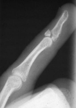
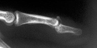
Posteroanterior (PA) and lateral radiographs centered at the distal interphalangeal (DIP) joint of the affected finger are required. These radiographs are used to differentiate between a bony mallet injury (see the first image below) and a tendinous one. They also reveal any associated metaphyseal, shaft, or tuft fractures of the distal phalanx. Perhaps most important, lateral radiographs reveal the presence of volar subluxation of the distal phalanx (see the second image below). In addition, these radiographic views reveal rare condylar fractures of the middle phalanx.
Radiographs of the whole hand do not suffice in the evaluation of mallet finger, because parallax of the x-ray beams creates an uninterpretable oblique view of the DIP joint. No imaging studies other than radiography are indicated in mallet finger.
-
Despite active extension effort, distal interphalangeal (DIP) joint of index finger rests in flexion, as is characteristic of mallet finger.
-
Typical mallet finger deformity.
-
Large dorsal-lip avulsion fracture from distal phalanx: bony mallet injury.
-
Mallet fracture with volar subluxation of distal phalanx.
-
Stable mallet fracture involving 40% of joint surface.
-
Dorsal aluminum foam splint for treatment of mallet finger.
-
Stack splints are widely used for treatment of mallet finger.
-
Molded plastic stack splint for treatment of mallet finger.
-
Skin-tight plaster cast can effectively hold distal interphalangeal (DIP) joint extended and proximal interphalangeal joint (PIP) flexed when mallet deformity is accompanied by hyperextensible PIP. Not immobilizing PIP in partial flexion risks development of swan-neck deformity.
-
Pressure injury can result from splint that is applied too tightly, especially if joint is maintained in hyperextended position rather than position of neutral extension.
-
Thermoplastic blank for custom-molded mallet finger splint; oblique view of molded splint in place.
-
Dorsal view of custom-molded thermoplastic splint in place.
-
Volar view of thermoplastic splint in place.
-
Application of thermoplastic splint.


