Approach Considerations
Charcot-Marie-Tooth (CMT) disease continues to be an incurable condition. Patients should be evaluated and treated symptomatically in a multidisciplinary approach by a team that includes the following [52] :
-
Neurologist
-
Physiatrist
-
Orthopedic surgeon
-
Physical therapist
-
Occupational therapist
-
Orthotists
-
Mental health provider
-
Genetic counselor
This approach is crucial for improving the quality of life of CMT patients. [53] Specialists in neurogenetics may be consulted to order specific genetic tests and proper genetic counseling.
Nonoperative Therapy
Currently, no proven medical treatment exists to reverse or slow the natural disease process for the underlying disorder. Nothing can correct the abnormal myelin, prevent its degeneration, or prevent axonal degeneration. [54] Improved understanding of the genetics and biochemistry of the disorder offers hope for an eventual treatment. Animal studies have suggested that targeting myelin lipid metabolism with lipid supplementation may be a potential therapeutic approach in CMT 1A. [55]
An insert with lateral posting and recession under the first ray can provide mechanical stability if the cavovarus deformity is flexible and correctible as tested with the Coleman block test. Additionally, the shoes can have a lateral flare along the outer border of the outsole, which can help in prevent the ankles from rolling over. High-top lace-up shoes can similarly provide additional stability.
A pilot study by Knak et al suggested that aerobic antigravity exercise may be helpful in patients with CMT 1A or CMT X; however, further evaluation in larger cohorts is needed. [56]
Surgical Therapy
Orthopedic surgery is required to correct severe pes cavus deformities, scoliosis, and other joint deformities. (See the images below.) Treatment is determined by the age of the patient and the cause and severity of the deformity.
Surgical procedures consist of the following three types:
-
Soft-tissue procedures (plantar fascia release, tendon release or transfer)
-
Osteotomy (metatarsal, midfoot, calcaneal)
-
Joint-stabilizing procedures (triple arthrodesis)
Procedures are usually staged. The initial procedure is a radical plantar or plantar-medial release-plantar fasciotomy, with a dorsal closing-wedge osteotomy of the first metatarsal base if necessary. Tendo calcaneus lengthening should not be performed as part of the initial procedure, because the force used to dorsiflex the forefoot causes the calcaneus to dorsiflex into an unacceptable position.
If the hindfoot is flexible and a posterior release is not necessary, posterior tibial tendon transfer can be done as part of the initial procedure for severe anterior tibial weakness. [57] In a prospective study of 14 patients with CMT disease who had cavovarus foot deformity, Dreher et al found that tibialis posterior tendon transfer was effective at correcting the foot-drop component of cavovarus foot deformity; the transfer apparently worked as an active substitution. [58]
When the hindfoot is flexible, early aggressive treatment with soft-tissue releases can delay the need for more extensive reconstructive procedures. The Jones procedure includes transfer of the extensor hallucis longus tendon to the first metatarsal head and arthrodesis of the interphalangeal (IP) joint of the great toe.
A review paper by Faldini et al concluded that plantar fasciotomy, midtarsal osteotomy, the Jones procedure, and dorsiflexion osteotomy of the first metatarsal yielded adequate correction of flexible cavus feet in patients with CMT disease in the absence of fixed hindfoot deformity. [59]
The Coleman block test (see the image below) is sometimes used to help decide what type of surgery is best. In cases of cavovarus deformity, this test evaluates hindfoot flexibility. [60] It is performed by placing the patient's foot on a wood block that is 2.5-4 cm thick, with the heel and lateral border of the foot on the block and bearing full weight while the first, second, and third metatarsals are allowed to hang freely into plantarflexion and pronation.
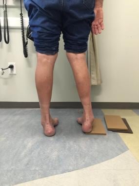 Coleman block test showing lack of correction of hindfoot varus malalignment when block is placed under outer border of foot.
If heel varus corrects while the patient is standing on the block, the hindfoot is considered flexible. If the subtalar joint is supple and corrects with the block test, then surgical procedures may be directed at correcting forefoot pronation, which is usually due to plantarflexion of the first metatarsal. If the hindfoot is rigid, then surgical correction of the forefoot and hindfoot is required.
Triple arthrodesis serves as a salvage procedure for patients in whom other procedures have been unsuccessful, as well as in patients with untreated fixed deformities.
Children younger than 8 years with supple hindfeet usually respond to plantar releases and appropriate tendon transfers. A first metatarsal osteotomy may be needed in some cases.
Children younger than 12 years with rigid hindfoot deformities may need radical plantar-medial release, first metatarsal osteotomy, and Dwyer lateral closing-wedge osteotomy of the calcaneus to correct the deformities.
In the early 1970s, the Akron dome osteotomy was developed as a salvage surgical option to manage rigid cavus deformity of the foot. In a retrospective study, Weiner et al showed that this operation is a valuable salvage procedure in the management of the rigid cavus deformity in children with CMT disease. [61]
Wukich and Bowen reported that only 14% of patients with CMT disease required triple arthrodesis. [62] They also reported hindfoot stability with triple arthrodesis, and when the posterior tibial tendon was transferred anteriorly, this eliminated the need for a postoperative drop-foot brace. They reported good or excellent results in 88% of patients who were treated with this method.
Ward et al studied the long-term results of surgical reconstruction procedures for cavovarus foot deformity in 25 patients with CMT disease who had undergone the procedure between 1970 and 1994 and were evaluated at a mean follow-up of 26.1 years. [63] The authors found that the use of soft-tissue procedures and first metatarsal osteotomy resulted in lower rates of degenerative changes and reoperations in comparison with results obtained with triple arthrodesis.
Generally, spinal deformities in children with CMT disease can be treated with the same techniques used for idiopathic scoliosis.
Prevention
Regular and proper follow-up and therapeutic interventions are necessary to avoid joint contractures and deformities.
Proper genetic counseling helps parents to understand the risk of having children with this disorder and gives them a chance to make informed decisions regarding pregnancy. [22, 36]
A study of mitochondrial data from 442 patients suggested that MT-ATP6 mutations are an important cause of CMT disease and can be evaluated with a simple blood test. [64]
Long-Term Monitoring
Patients should have regular follow-up visits to check for deterioration in function and the development of contractures. This follow-up allows early detection of complications. Proper interventions early in the disease course help to avoid significant and permanent functional limitations. [37]
-
Foot deformities in 16-year-old boy with Charcot-Marie-Tooth disease type 1A.
-
Charcot-Marie-Tooth disease type 1A DNA test showing duplication in short arm of chromosome 17 (A); compared with normal (B).
-
Nerve conduction study showing decreased nerve conduction velocity in median nerve in 18-year-old woman with Charcot-Marie-Tooth disease type 1.
-
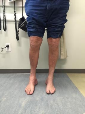
Cavovarus feet with heels visible from front ("peekaboo" sign).
-
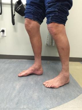
High arch typical of patients with cavus feet.
-
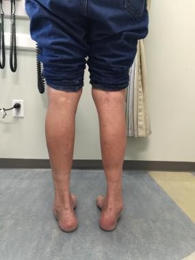
Both heels showing varus deformity when observed from back.
-
Coleman block test showing lack of correction of hindfoot varus malalignment when block is placed under outer border of foot.
Tables
CMT Type |
Chromosome; Inheritance Pattern |
Age of Onset |
Clinical Features |
Average NCVs§ |
CMT 1A (PMP-22¶ dupl.) |
17p11; AD* |
First decade |
Distal weakness |
15-20 m/s |
CMT 1B (P0 -MPZ)** |
1q22; AD |
First decade |
Distal weakness |
< 20 m/s |
CMT 1C (non A, non B) |
16p13;AD |
Second decade |
Distal weakness |
26-42 m/s |
10q21; AD |
First decade |
Distal weakness |
15-20 m/s |
|
CMT 1E |
17p11; AD |
First decade |
Distal weakness, deafness |
15-20 m/s |
CMT 1F |
8p21; AD |
First decade |
Distal weakness |
15-20 m/s |
Xq13; XD‡ |
Second decade |
Distal weakness |
25-40 m/s |
|
CMT 2A |
1p36; AD |
10 y |
Distal weakness |
>38 m/s |
CMT 2B |
3q; AD |
Second decade |
Distal weakness, sensory loss, skin ulcers |
Axon loss; Normal |
CMT 2C |
12q23-q24, AD |
First decade |
Vocal cord, diaphragm, and distal weakness |
>50 m/s |
CMT 2D |
7p14; AD |
16-30 y |
Distal weakness, upper limb predominantly |
Axon loss; N†† |
CMT 2E |
8p21; AD |
10-30 y |
Distal weakness, lower limb predominantly |
Axon loss; N |
CMT 2F |
7q11-q21; AD |
15-25 y |
Distal weakness |
Axon loss; N |
CMT 2G |
12q12-q13; ?AD |
9-76 y |
Distal weakness |
Axon loss; N |
CMT 2H |
?; AR† |
15-25 y |
Distal weakness, pyramidal features |
Axon loss; N |
CMT 2I |
1q22; AD |
47-60 y |
Distal weakness |
Axon loss; N |
CMT 2J |
1q22; AD |
40-50 y |
Distal weakness, hearing loss |
Axon loss; N |
CMT 2K |
8q13-q21; AR |
< 4 y |
Distal weakness |
Axon loss; N |
CMT 2L |
12q24; AD |
15-25 y |
Distal weakness |
Axon loss; N |
CMT R-Ax (Ouvrier) |
AR |
First decade |
Distal weakness |
Axon loss; N |
CMT R-Ax (Moroccan) |
1q21; AR |
Second decade |
Distal weakness |
Axon loss; N |
Cowchock syndrome |
Xq24-q26 |
First decade |
Distal weakness, deafness, intellectual disability |
Axon loss; N |
HNPP|| (PMP-22) Or tomaculous neuropathy |
17p11; AD |
All ages |
Episodic weakness and numbness |
Conduction Blocks |
Dejerine-Sottas syndrome (DSS) or hereditary motor and sensory neuropathy (HMSN) 3 |
P0; AR PMP-22; AD 8q23; AD |
2 y |
Severe weakness |
< 10 m/s |
Congenital hypomyelination (CH) |
P0, EGR2 or PMP-22 AR |
Birth |
Severe weakness |
< 10 m/s |
CMT 4A |
8q13; AR |
Childhood |
Distal weakness |
Slow |
CMT 4B (Myotubular in-related protein-2) [18] |
11q23; AR |
2-4 y |
Distal and proximal weakness |
Slow |
CMT 4C |
5q23; AR |
5-15 y |
Delayed walking |
14-32 m/s |
CMT 4D (Lom) (N-myc Downstream- Regulated Gene 1) |
8q24; AR |
1-10 y |
Distal muscle wasting, foot and hand deformities |
10-20 m/s |
CMT 4E (EGR2) |
10q21; AR |
Birth |
Infant hypotonia |
9-20 m/s |
CMT 4G |
10q23.2; AR |
8-16 years |
Distal weakness |
9-20 m/s |
CMT 4H |
12p11.21-q13.11; AR |
0-2 years |
Delayed walking |
9-20 m/s |
CMT 4F |
19q13; AR |
1-3 y |
Motor delay |
Absent |
*Autosomal dominant †Autosomal recessive ‡X-linked dominant §Nerve conduction velocities ||Hereditary neuropathy with liability to pressure palsy ¶Peripheral myelin protein #Early growth response **Myelin protein zero ††Normal |
||||