Frieden IJ. Aplasia cutis congenita: a clinical review and proposal for classification. J Am Acad Dermatol. 1986 Apr. 14(4):646-60. [QxMD MEDLINE Link].
Caksen H, Kurtoglu S. Our experience with aplasia cutis congenita. J Dermatol. 2002 Jun. 29(6):376-9. [QxMD MEDLINE Link].
Moros Pena M, Labay Matias M, Valle Sanchez F, et al. [Aplasia cutis congenita in a newborn: etiopathogenic review and diagnostic approach]. An Esp Pediatr. 2000 May. 52(5):453-6. [QxMD MEDLINE Link].
Vinay K, Yadav S, Parsad D, Abhijit C. Aplasia cutis congenita in a Blaschkoid distribution: a lesser known variant. Int J Dermatol. 2016 Jan 11. [QxMD MEDLINE Link].
Hennekam RC. Aplasia cutis congenita reminiscent of the lines of Blaschko. Hum Genet. 1992 Dec. 90 (4):469-71. [QxMD MEDLINE Link].
Rokunohe D, Akasaka E, Rokunohe A, Kaneko T, Matsuzaki Y, Takiyoshi N, et al. Multiple aplasia cutis congenita lesions located along Blaschko's lines in a patient with tetralogy of Fallot-A. J Dermatol Case Rep. 2012 Jun 30. 6 (2):40-2. [QxMD MEDLINE Link].
Drolet B, Prendiville J, Golden J, Enjolras O, Esterly NB. Membranous aplasia cutis' with hair collars. Congenital absence of skin or neuroectodermal defect?. Arch Dermatol. 1995 Dec. 131(12):1427-31. [QxMD MEDLINE Link].
Colon-Fontanez F, Fallon Friedlander S, Newbury R, Eichenfield LF. Bullous aplasia cutis congenita. J Am Acad Dermatol. 2003 May. 48(5 Suppl):S95-8. [QxMD MEDLINE Link].
Benjamin LT, Trowers AB, Schachner LA. Giant aplasia cutis congenita without associated anomalies. Pediatr Dermatol. 2004 Mar-Apr. 21(2):150-3. [QxMD MEDLINE Link].
Chitnis MR, Carachi R, Galea P. Familial aplasia cutis congenita. Eur J Pediatr Surg. 1996 Apr. 6(2):100-1. [QxMD MEDLINE Link].
Fagan LL, Harris PA, Coran AG, Cywes R. Sporadic aplasia cutis congenita. Pediatr Surg Int. 2002 Sep. 18(5-6):545-7. [QxMD MEDLINE Link].
Davidson AW, Hosalkar HS, Hill RA, Monsell F. Radial dysplasia with localized cutis aplasia congenita. J Pediatr Orthop B. 2003 Nov. 12(6):398-401. [QxMD MEDLINE Link].
Balasubramanian M, Collins AL. Aplasia cutis congenita, terminal limb defects and periventricular leukomalacia in one sibling with minor findings in the other-probable autosomal recessive Adams-Oliver Syndrome. Eur J Med Genet. 2009 Jul-Aug. 52(4):234-8. [QxMD MEDLINE Link].
Bilginer B, Onal MB, Bahadir S, Akalan N. Aplasia cutis congenita of the scalp, skull and dura associated with Adams-Oliver syndrome. Turk Neurosurg. 2008 Apr. 18(2):191-3. [QxMD MEDLINE Link].
Dyall-Smith D, Ramsden A, Laurie S. Adams-Oliver syndrome: aplasia cutis congenita, terminal transverse limb defects and cutis marmorata telangiectatica congenita. Australas J Dermatol. 1994. 35(1):19-22. [QxMD MEDLINE Link].
Orstavik KH, Stromme P, Spetalen S, et al. Aplasia cutis congenita associated with limb, eye, and brain anomalies in sibs: a variant of the Adams-Oliver syndrome?. Am J Med Genet. 1995 Oct 23. 59(1):92-5. [QxMD MEDLINE Link].
Temtamy SA, Aglan MS, Ashour AM, Zaki MS. Adams-Oliver syndrome: further evidence of an autosomal recessive variant. Clin Dysmorphol. 2007 Jul. 16(3):141-9. [QxMD MEDLINE Link].
Meester JA, Southgate L, Stittrich AB, Venselaar H, Beekmans SJ, den Hollander N, et al. Heterozygous Loss-of-Function Mutations in DLL4 Cause Adams-Oliver Syndrome. Am J Hum Genet. 2015 Sep 3. 97 (3):475-82. [QxMD MEDLINE Link].
Hassed SJ, Wiley GB, Wang S, Lee JY, Li S, Xu W, et al. RBPJ mutations identified in two families affected by Adams-Oliver syndrome. Am J Hum Genet. 2012 Aug 10. 91 (2):391-5. [QxMD MEDLINE Link].
Shaheen R, Aglan M, Keppler-Noreuil K, Faqeih E, Ansari S, Horton K, et al. Mutations in EOGT confirm the genetic heterogeneity of autosomal-recessive Adams-Oliver syndrome. Am J Hum Genet. 2013 Apr 4. 92 (4):598-604. [QxMD MEDLINE Link].
Shaheen R, Faqeih E, Sunker A, Morsy H, Al-Sheddi T, Shamseldin HE, et al. Recessive mutations in DOCK6, encoding the guanidine nucleotide exchange factor DOCK6, lead to abnormal actin cytoskeleton organization and Adams-Oliver syndrome. Am J Hum Genet. 2011 Aug 12. 89 (2):328-33. [QxMD MEDLINE Link].
Southgate L, Machado RD, Snape KM, Primeau M, Dafou D, Ruddy DM, et al. Gain-of-function mutations of ARHGAP31, a Cdc42/Rac1 GTPase regulator, cause syndromic cutis aplasia and limb anomalies. Am J Hum Genet. 2011 May 13. 88 (5):574-85. [QxMD MEDLINE Link].
Stittrich AB, Lehman A, Bodian DL, Ashworth J, Zong Z, Li H, et al. Mutations in NOTCH1 cause Adams-Oliver syndrome. Am J Hum Genet. 2014 Sep 4. 95 (3):275-84. [QxMD MEDLINE Link].
Narang T, Kanwar AJ, Dogra S. Adams-Oliver syndrome: a sporadic occurrence with minimal disease expression. Pediatr Dermatol. 2008 Jan-Feb. 25(1):115-6. [QxMD MEDLINE Link].
Lam J, Dohil MA, Eichenfield LF, Cunningham BB. SCALP syndrome: sebaceous nevus syndrome, CNS malformations, aplasia cutis congenita, limbal dermoid, and pigmented nevus (giant congenital melanocytic nevus) with neurocutaneous melanosis: a distinct syndromic entity. J Am Acad Dermatol. 2008 May. 58(5):884-8. [QxMD MEDLINE Link].
Neri I, Savoia F, Giacomini F, Raone B, Aprile S, Patrizi A. Usefulness of dermatoscopy for the early diagnosis of sebaceous naevus and differentiation from aplasia cutis congenita. Clin Exp Dermatol. 2009 Jul. 34(5):e50-2. [QxMD MEDLINE Link].
Kantor J, Yan AC, Hivnor CM, Honig PJ, Kirschner R. Extensive aplasia cutis congenita and the risk of sagittal sinus thrombosis. Arch Dermatol. 2005 May. 141(5):554-6. [QxMD MEDLINE Link].
Kim CS, Tatum SA, Rodziewicz G. Scalp aplasia cutis congenita presenting with sagittal sinus hemorrhage. Arch Otolaryngol Head Neck Surg. 2001 Jan. 127(1):71-4. [QxMD MEDLINE Link].
Lane W, Zanol K. Duodenal atresia, biliary atresia, and intestinal infarct in truncal aplasia cutis congenita. Pediatr Dermatol. 2000 Jul-Aug. 17(4):290-2. [QxMD MEDLINE Link].
Ribuffo D, Costantini M, Gullo P, Houseman ND, Taylor GI. Aplasia cutis congenita of the scalp, the skull, and the dura. Scand J Plast Reconstr Surg Hand Surg. 2003. 37(3):176-80. [QxMD MEDLINE Link].
Gómez M, Chiesura V, Noguera-Morel L, Hernández-Martín A, García-Peñas JJ, Torrelo A. Extensive Intracranial Arteriovenous Malformation in a Child with Aplasia Cutis Congenita. Pediatr Dermatol. 2015 Jul-Aug. 32 (4):e163-4. [QxMD MEDLINE Link].
Schierz IAM, Giuffrè M, Del Vecchio A, Antona V, Corsello G, Piro E. Recognizable neonatal clinical features of aplasia cutis congenita. Ital J Pediatr. 2020 Feb 18. 46 (1):25. [QxMD MEDLINE Link].
Kelly BJ, Samolitis NJ, Xie DL, Skidmore RA. Aplasia cutis congenita of the trunk with fetus papyraceus. Pediatr Dermatol. 2002 Jul-Aug. 19(4):326-9. [QxMD MEDLINE Link].
Maccario S, Fasolato V, Brunelli A, Martinelli S. Aplasia cutis congenita: an association with vanishing twin syndrome. Eur J Dermatol. 2009 Jul-Aug. 19(4):372-4. [QxMD MEDLINE Link].
Schaffer JV, Popiolek DA, Orlow SJ. Symmetric truncal aplasia cutis congenita following multifetal reduction of a sextuplet pregnancy. J Pediatr. 2008 Dec. 153(6):860-3. [QxMD MEDLINE Link].
Simman R. Reconstruction aplasia cutis congenita (group V) of the trunk in a newborn. Plast Reconstr Surg. 2004 Mar. 113(3):1103. [QxMD MEDLINE Link].
Visva-Lingam S, Jana A, Murray H, John E. Preterm premature rupture of membranes associated with aplasia cutis congenita and fetus papyraceous. Aust N Z J Obstet Gynaecol. 1996 Feb. 36(1):90-1. [QxMD MEDLINE Link].
Pieretti ML, Alcalá R, Boggio P, Noguera-Morel L, Porriño ML, Luna PC, et al. Aplasia Cutis Congenita Associated with Fetus Papyraceus. Pediatr Dermatol. 2015 Nov-Dec. 32 (6):858-61. [QxMD MEDLINE Link].
Chan RK, Liu AS, Rogers GF. Aplasia cutis congenita of the trunk associated with fetus papyraceous. J Craniofac Surg. 2012 Jul. 23(4):995-7. [QxMD MEDLINE Link].
McCarthy MA, Clarke T, Powell FC. Epidermolysis bullosa and aplasia cutis. Int J Dermatol. 1991 Jul. 30(7):481-4. [QxMD MEDLINE Link].
Bigliardi PL, Braschler C, Kuhn P, Sigrist J, Buechner S, Rufli T. Unilateral aplasia cutis congenita on the leg. Pediatr Dermatol. 2004 Jul-Aug. 21(4):454-7. [QxMD MEDLINE Link].
Benvenuto C, Kraemer CK, Kruse RL, Cestari TF. Familial epidermolysis bullosa with aplasia cutis congenita: Bart's syndrome?. Skinmed. 2003 Sep-Oct. 2(5):319-21. [QxMD MEDLINE Link].
Atik B, Tan O, Bayram I, Tuncer O, Kirimi E. Asymmetrical nonscalp aplasia cutis congenita: a case report. J Dermatol. 2004 Nov. 31(11):923-6. [QxMD MEDLINE Link].
Boente Mdel C, Frontini Mdel V, Acosta MI, Saleme C, Barrionuevo S, Asial R. Extensive symmetric truncal aplasia cutis congenita without fetus papyraceus or macroscopic evidence of placental abnormalities. Pediatr Dermatol. 1995 Sep. 12(3):228-30. [QxMD MEDLINE Link].
Morrell DS, Rubenstein DS, Briggaman RA, Fine JD, Pulkkinen L, Uitto J. Congenital pyloric atresia in a newborn with extensive aplasia cutis congenita and epidermolysis bullosa simplex. Br J Dermatol. 2000 Dec. 143(6):1342-3. [QxMD MEDLINE Link].
Izhar R, Ghani T. Aplasia cutis congenita and antithyroid drugs. J Pak Med Assoc. 2002 Nov. 52(11):526-8. [QxMD MEDLINE Link].
Karg E, Bereg E, Gaspar L, Katona M, Turi S. Aplasia cutis congenita after methimazole exposure in utero. Pediatr Dermatol. 2004 Jul-Aug. 21(4):491-4. [QxMD MEDLINE Link].
Mandel SJ, Brent GA, Larsen PR. Review of antithyroid drug use during pregnancy and report of a case of aplasia cutis. Thyroid. 1994 Spring. 4(1):129-33. [QxMD MEDLINE Link].
Nakamura S, Nishikawa T, Isaji M, et al. Aplasia cutis congenita and skull defects after exposure to methimazole in utero. Intern Med. 2005 Nov. 44(11):1202-3. [QxMD MEDLINE Link].
Evers ME, Steijlen PM, Hamel BC. Aplasia cutis congenita and associated disorders: an update. Clin Genet. 1995 Jun. 47(6):295-301. [QxMD MEDLINE Link].
Khan JY, Moss C, Roper HP. Aplasia cutis congenita with chromosome 12q abnormality. Arch Dis Child Fetal Neonatal Ed. 1995 May. 72(3):F205-6. [QxMD MEDLINE Link]. [Full Text].
Casanova D, Amar E, Bardot J, Magalon G. Aplasia cutis congenita. Report on 5 family cases involving the scalp. Eur J Pediatr Surg. 2001 Aug. 11(4):280-4. [QxMD MEDLINE Link].
Rodrigues RG. Aplasia cutis congenita, congenital heart lesions, and frontonasal cysts in four successive generations. Clin Genet. 2007 Jun. 71(6):558-60. [QxMD MEDLINE Link].
Teebi AS, Druker HA. Brachycephaly, cutis aplasia congenita, blue sclerae, hypertelorism, polydactyly, hypoplastic nipples, failure to thrive, and developmental delay: a distinct autosomal recessive syndrome?. Clin Dysmorphol. 2001 Jan. 10(1):69-70. [QxMD MEDLINE Link].
Sugiura T, Kouwaki M, Kiyosawa S, et al. A case of systemic aplasia cutis congenita: a newly recognized syndrome?. Eur J Pediatr. 2008 Apr. 167(4):409-13. [QxMD MEDLINE Link].
Marneros AG, Beck AE, Turner EH, McMillin MJ, Edwards MJ, Field M, et al. Mutations in KCTD1 cause scalp-ear-nipple syndrome. Am J Hum Genet. 2013 Apr 4. 92 (4):621-6. [QxMD MEDLINE Link].
Canham NL. Cutis aplasia as a feature of Kabuki syndrome. Clin Dysmorphol. 2006 Jul. 15(3):179-80. [QxMD MEDLINE Link].
Marneros AG. BMS1 is mutated in aplasia cutis congenita. PLoS Genet. 2013 Jun. 9(6):e1003573. [QxMD MEDLINE Link]. [Full Text].
Baselga E, Torrelo A, Drolet BA, Zambrano A, Alomar A, Esterly NB. Familial nonmembranous aplasia cutis of the scalp. Pediatr Dermatol. 2005 May-Jun. 22(3):213-7. [QxMD MEDLINE Link].
Martinez-Regueira S, Vazquez-Lopez ME, Somoza-Rubio C, Morales-Redondo R, Gonzalez-Gay MA. Aplasia cutis congenita in a defined population from northwest Spain. Pediatr Dermatol. 2006 Nov-Dec. 23 (6):528-32. [QxMD MEDLINE Link].
Tempark T, Shwayder TA. Aplasia cutis congenita with fetus papyraceus: report and review of the literature. Int J Dermatol. 2012 Dec. 51 (12):1419-26. [QxMD MEDLINE Link].
Gerber M, de Veciana M, Towers CV, Devore GR. Aplasia cutis congenita: a rare cause of elevated alpha-fetoprotein levels. Am J Obstet Gynecol. 1995 Mar. 172(3):1040-1. [QxMD MEDLINE Link].
Drolet BA, Clowry L Jr, McTigue MK, Esterly NB. The hair collar sign: marker for cranial dysraphism. Pediatrics. 1995 Aug. 96(2 Pt 1):309-13. [QxMD MEDLINE Link].
Browning JC. Aplasia cutis congenita: approach to evaluation and management. Dermatol Ther. 2013 Nov. 26(6):439-44. [QxMD MEDLINE Link].
Humphrey SR, Hu X, Adamson K, Schaus A, Jensen JN, Drolet B. A practical approach to the evaluation and treatment of an infant with aplasia cutis congenita. J Perinatol. 2018 Feb. 38 (2):110-117. [QxMD MEDLINE Link].
Santos de Oliveira R, Barros Juca CE, Lopes Lins-Neto A, Aparecida do Carmo Rego M, Farina J, Machado HR. Aplasia cutis congenita of the scalp: is there a better treatment strategy?. Childs Nerv Syst. 2006 Sep. 22(9):1072-9. [QxMD MEDLINE Link].
Gan YC, Steinbok P. Aplasia cutis congenita of the scalp: is there a better treatment strategy?. Childs Nerv Syst. 2006 Oct. 22(10):1216-7; author reply 1218-9. [QxMD MEDLINE Link].
Ahcan U, Janezic T. Management of aplasia cutis congenita in a non-scalp location. Br J Plast Surg. 2002 Sep. 55(6):530-2. [QxMD MEDLINE Link].
Bang RL, Ghoneim IE, Gang RK, Al Najjadah I. Treatment dilemma: conservative versus surgery in cutis aplasia congenita. Eur J Pediatr Surg. 2003 Apr. 13(2):125-9. [QxMD MEDLINE Link].
Donati V, Arena S, Capilli G, Carrera G, Ciralli F, Liberatore A. Reparation of a severe case of aplasia cutis congenita with engineered skin. Biol Neonate. 2001. 80(4):273-6. [QxMD MEDLINE Link].
Madsen JR, Robertson RL, Bartlett R. Surgical management of cutis aplasia with high-flow sinus pericranii. Pediatr Neurosurg. 1998 Feb. 28(2):79-83. [QxMD MEDLINE Link].
Rhee ST, Colville C, Buchman SR, Muraszko K. Complete osseous regeneration of a large skull defect in a patient with cutis aplasia: a conservative approach. J Craniofac Surg. 2002 Jul. 13(4):497-500. [QxMD MEDLINE Link].
Shivakumar SK, Dwarakanath S, Swaroop G, Venkataramana NK. Aplasia cutis congenita of the scalp: therapeutic modalities. Neurol India. 2006 Sep. 54(3):312-3. [QxMD MEDLINE Link].
Skoufi G, Lialios G, Plachouras N, Kutsogiannis D, Mperis A. Aplasia cutis congenita: Successful conservative treatment. Pediatr Int. 2006 Oct. 48(5):507-9. [QxMD MEDLINE Link].
Maillet-Declerck M, Vinchon M, Guerreschi P, Pasquesoone L, Dhellemmes P, Duquennoy-Martinot V, et al. Aplasia Cutis Congenita: Review of 29 Cases and Proposal of a Therapeutic Strategy. Eur J Pediatr Surg. 2012 Aug 17. [QxMD MEDLINE Link].
Azad S, Falder S, Harrison J, Graham K. An adherent dressing for aplasia cutis congenita. Br J Plast Surg. 2005 Dec. 58(8):1159-61. [QxMD MEDLINE Link].
Lahiri A, Nishikawa H. A nonadherent dressing for aplasia cutis congenita. J Plast Reconstr Aesthet Surg. 2006. 59(7):781-2. [QxMD MEDLINE Link].
Orgun D, Horiguchi M, Hayashi A, Shimoji K, Arai H, Mizuno H. Conservative Treatment of Large Aplasia Cutis Congenita of the Scalp With Bone Defect With Basic Fibroblast Growth Factor Application. J Craniofac Surg. 2017 Mar. 28 (2):e154-e158. [QxMD MEDLINE Link].
Mericli AF, Chen K, Murariu D, Jane JA Jr, Lin KY. Calvarial Regeneration With Use of Acellular Dermal Matrix in Aplasia Cutis Congenita. J Craniofac Surg. 2015 Sep. 26 (6):1960-2. [QxMD MEDLINE Link].
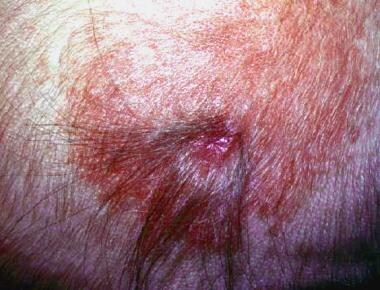 This area of healed aplasia cutis congenita is located in an area of nevus flammeus. Note the collarette of coarser hair at the margin of the defect.
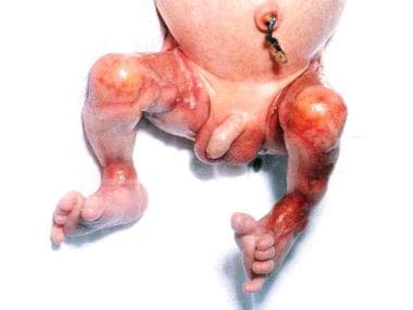 Bilateral involvement of the lower extremities in aplasia cutis congenita associated with fetus papyraceous.


