Practice Essentials
An ectopic pregnancy is any non-intrauterine pregnancy (IUP) and can be classified as tubal, interstitial, ovarian, abdominal, heterotopic, cervical, and cesarean scar. The most common location is the fallopian tubes. [1] Clinicians should consider the diagnosis of ectopic pregnancy in any woman in the first trimester of pregnancy who has abdominal or pelvic pain, vaginal bleeding, or both. [2, 3, 4] The incidence of ectopic pregnancy is 1-2% and is still the most common cause of pregnancy-related death in the first trimester, accounting for about 10% of all pregnancy-related deaths. Missed ectopic pregnancy is a leading cause of emergency medicine malpractice claims. [5, 6, 7, 8]
High-risk features for the possibility of ectopic pregnancy include history of ectopic pregnancy, history of pelvic inflammatory disease, use of an intrauterine device, and history of tubal surgery. Unfortunately, clinical findings alone cannot reliably diagnose or exclude ectopic pregnancy. [9, 10, 11]
Bedside ultrasonography is indicated in the presence of vaginal bleeding or abdominal pain in a patient in the first trimester of pregnancy. Do not perform bedside ultrasonography if it delays resuscitation or definitive surgical care in an unstable patient.
Several studies have validated pelvic ultrasonography in the acute care setting, specifically in the emergency department (ED), as diagnostically accurate and beneficial for flow. [12, 13, 14, 15, 16, 17, 18]
One meta-analysis of emergency physician–performed ultrasonography as a diagnostic test for ectopic pregnancy found that it had a sensitivity of 99.3% and a negative predictive value of 99.96% for detecting an intrauterine pregnancy. Given a disease prevalence of 7.5% and a negative likelihood ratio of 0.08, visualization of intrauterine pregnancy by an emergency physician yields a posttest probability of ectopic pregnancy of 0.6%. [19] A retrospective study of 585 women over a 2.5-year period concluded that the sensitivity and specificity of ultrasound for the detection of ectopic pregnancy was 88.5% and 96.5% on the first ultrasound and 93.1% and 95.7% after an additional scan. [20]
An early diagnosis of ruptured ectopic pregnancy is necessary, because it is the leading cause of first-trimester maternal death. There have also been reports of an increase in mortality in patients with late post-hysterectomy ectopic pregnancy. [21]
A study by Stone et al revealed that compared to radiology department–performed ultrasound (RADUS) alone, incorporation of point-of care ultrasound (POCUS) was associated with significantly faster ED treatment time for all ectopic pregnancies and significantly faster time to the operating room (OR) for ruptured ectopic pregnancies, even when combined with RADUS. The average ED treatment time in the POCUS group for all admitted ectopic pregnancies was 157.9 minutes versus 206.3 minutes in the RADUS group. The median time to OR for ruptured ectopic pregnancies was 203.0 minutes in the POCUS group versus 293.0 min in the RADUS group. [22]
Urquhart et al found that ED patients with a ruptured ectopic pregnancy who first received POCUS had shorter times to diagnosis, obstetric consultation, and/or arrival as compared to those who received RADUS. Comparing times between POCUS and RADUS groups, the mean time from ED arrival to ultrasound interpretation was 15 minutes versus 138 minutes, from ED arrival to obstetric consultation was 35 minutes versus 150 minutes, from ED arrival to OR arrival was 160 minutes versus 381 minutes, and from ultrasound interpretation to OR arrival was 145 minutes versus 243 minutes. [23]
In a first-trimester study of ultrasound features for diagnosis of ectopic pregnancy, an empty uterus was found to predict an ectopic pregnancy with a sensitivity of 81.1% and a specificity of 79.5%. Sensitivity and specificity for a pseudosac, adnexal mass, and free fluid were as follows: 5.5% and 94.2%; 63.5% and 91.4%; and 47.2% and 92.3%, respectively. [24]
Bedside ultrasonography is an important tool for emergency medicine clinicians and other acute care clinicians to use in assessing patients’ risk for potential ectopic pregnancy. Early diagnosis can be very valuable in lessening morbidity and mortality. Diagnosis before tubal rupture can prevent life-threatening hemorrhage and increase the probability that the patient may be managed medically or via tube-conserving surgery.
However, use of ultrasonographic imaging should never preclude adequate resuscitation or definitive surgical therapy in a patient who is hemodynamically unstable and in whom ectopic pregnancy is strongly suspected.
Ectopic pregnancy can usually be reliably excluded in patients with a demonstrated IUP; heterotopic pregnancy remains very rare in patients who are not taking fertility agents. Risk factors for heterotopic pregnancy include a family history of multiple gestations, endometriosis, tubal disease, pelvic inflammation, and high female hormone levels. The risk also increases with higher numbers of transferred embryos. Heterotopic pregnancies occur in approximately 1 in 5,000 pregnancies, but the incidence increases to as high as 1 in 100 in women undergoing fertility stimulation or procedures. [25]
This limited diagnostic focus differs from that of the ultrasonography performed by the radiology department and has also been called point-of-care limited ultrasonography (PLUS). When the serum level of beta–human chorionic gonadotropin (β-hCG) is higher than 1500 mIU/mL, the level known as the discriminatory zone, transvaginal ultrasonographic findings of an IUP should be present (see the image below).
Diagnostic, Suggestive, and Indeterminate Ultrasonographic Findings
The first developmental structure big enough to be visualized by transvaginal ultrasonography is the gestational sac, which appears in the endometrial cavity at around 4.5-5 weeks’ gestation (corresponding to a β-HCG level of 1000-1500 mIU/mL). Measurement of the mean sac diameter (MSD) is important for estimating the gestational age, as well as for confirming subsequent normal embryonic development. [26]
According to a study by Oh et al, there was no difference in gestational sac diameter at 28-35 days after the last menstrual period in normal and abnormal pregnancies, but smaller-than-expected sac diameter in pregnancies 36-42 days after the last menstrual period was found to be predictive of spontaneous miscarriage. [27]
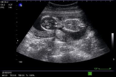
A conservative definition of a sonogram diagnostic for an IUP involves demonstration of a clearly defined yolk sac within the gestational sac (see the images below).
The yolk sac appears by 5-6 weeks’ gestation and should definitely be present when the MSD is greater than 8 mm. The embryo, or fetal pole, can be visualized on transvaginal ultrasonography by 6 weeks’ gestation and on transabdominal ultrasonography by 7 weeks’ gestation, and it should be present when the MSD exceeds 16 mm.
Correlated Sonographic Findings with Gestational Age (Open Table in a new window)
| Sonographic Finding | Gestational Age (weeks) Identified by Transvaginal Ultrasound | Gestational Age (weeks) Identified by Transabdominal Ultrasound |
|---|---|---|
| Gestational Sac | 4.5 - 5 | Unable to visualize |
| Yolk Sac | 5 - 6 | 6 - 7 |
| Fetal Pole | 6 | 7 |
Studies have shown that a cutoff of 25 mm can increase sensitivity to 100%. [28]
Embryonic cardiac activity starts to be visible at around 7 weeks’ gestation and should be visible if the crown-rump length, or fetal pole length, is greater than 5 mm. [29, 30]
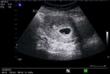
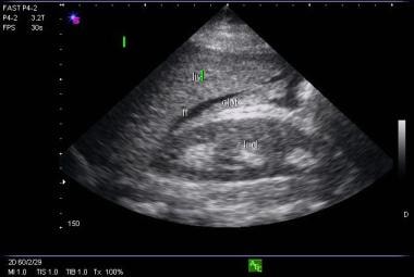
Definitive ultrasonographic diagnosis of an ectopic pregnancy is made in only about 20% of cases, when an extrauterine pregnancy is clearly identified (ie, an extrauterine gestational sac with a yolk sac or fetal pole is visualized). There exist, however, numerous findings that are highly suggestive of ectopic pregnancy, including an empty uterus in a patient with a β-hCG level above the discriminatory zone, an adnexal mass other than a simple cyst (see the image below), echogenic fluid in the cul-de-sac, or anything more than a small amount of fluid in the cul-de-sac.
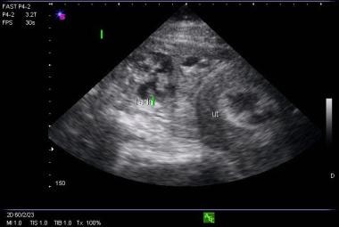 Picture of uterus without a fetal pole and a complex adnexal mass consistent with ectopic pregnancy.
Patients who exhibit such findings should be managed in consultation with an obstetrician; they likely will need surgical exploration or medical therapy with methotrexate.
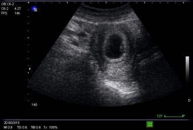
Ultrasonographic findings that are neither diagnostic nor highly suggestive of an IUP or ectopic pregnancy are classified as indeterminate. These findings include an empty uterus, an abnormal gestational sac (eg, a sac with an irregular border or an MSD large enough that a secondary structure such as a yolk sac would be expected), a normal gestational sac without a yolk sac or embryo, a nonspecific intrauterine fluid collection, and ill-defined echogenic material within the endometrial cavity (see the image below). Patients who exhibit these findings are generally monitored closely with serial β-HCG testing and clinical assessments, as about 10-25% of such patients have normal pregnancies.
Complications
There are no major complications associated with the performance of emergency department (ED) ultrasonography to evaluate first-trimester pregnancy. Sometimes, Doppler ultrasonography is used in early pregnancy evaluation to detect fetal cardiac activity or to better delineate the adjacent vascular anatomy. The energy output of Doppler ultrasonography is substantially higher than that of conventional ultrasonography and may be harmful to the embryo. [31]
Complications can ensue if an obstetrician is not consulted early in the treatment of a patient with early pregnancy and hemodynamic instability, acute abdomen, or falling hematocrit level.
Preparation
Anesthesia is generally not necessary for sonographic evaluation. However, patients may experience discomfort from the pressure of the transducer. Equipment includes the following:
-
Ultrasonograph with 3.5-5 MHz abdominal transducer and 7.5-10 MHz transvaginal transducer
-
Gloves
-
Acoustic gel
-
Transvaginal transducer probe cover
-
Lubricating jelly: Because acoustic gel causes an intravaginal dermatitis, the lubricating jelly Surgilube should be used on the outside of the transducer cover.
The patient should be in a hospital gown, undressed from the waist down. For transabdominal ultrasonography, position the patient in the supine recumbent position. For transvaginal ultrasonography, position the patient in the supine lithotomy position.
Be sure to scan systematically and widely, including the entire uterus and cervix in the transverse and sagittal planes. Failure to scan through the entire uterus, cervix, and adnexa can lead to missed ectopic pregnancies.
Explain the procedure, benefits, risks, and complications to the patient or the patient’s representative. Ask the patient or the patient’s representative if he or she would like others to be present for the procedure.
Technique
Overview
The primary role of emergency department (ED) obstetric ultrasonography is to demonstrate an intrauterine pregnancy (IUP). When the beta–human chorionic gonadotropin (β-hCG) level is greater than 1500 mIU/mL, there should be transvaginal ultrasonographic evidence of an IUP. The first visible structure is the gestational sac. Subsequent yolk sac, embryo, and fetal cardiac activity should appear at predictable time intervals and mean sac diameter sizes.
An extrauterine gestational sac with yolk sac or fetal pole is definitive evidence of an ectopic pregnancy. (Note that a pseudogestational sac can be confused with a genuine gestational sac.) Other suggestive ultrasonographic findings include a complex adnexal mass, echogenic fluid in the cul-de-sac, a moderate to large amount of fluid in the cul-de-sac, and an empty uterus in a patient with a β-HCG level above the discriminatory zone. (Note also that intrauterine fluid is one of the most common ectopic findings misinterpreted as an early normal IUP.)
Transabdominal ultrasonography
Transabdominal ultrasonography is best performed on a patient who has a full bladder.
Expose the abdomen from xiphoid to pubis. Apply a generous amount of acoustic gel to the patient’s lower abdomen, the abdominal transducer, or both.
The uterus is a muscular, hollow organ behind the bladder and anterior to the colon, with a moderately echogenic, homogeneous myometrium and a relatively hyperechoic endometrium.
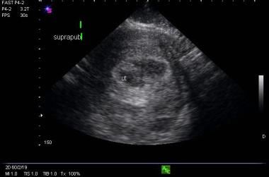
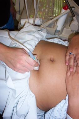
First, scan the uterus in the transverse plane. Hold the probe perpendicular to the patient’s long axis, with the indicator of the probe pointing toward the patient’s right, and sweep from fundus to cervix (see the image below).
Next, scan the uterus in the sagittal plane. Hold the probe parallel to the patient’s long axis, with the indicator of the probe pointing toward the patient’s head, and sweep from side to side (see the images below). Be sure to identify the landmarks: the bladder, the vaginal stripe, and the uterus.
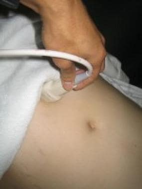 Sagittal or longitudinal probe placement for transabdominal obstetric examination.
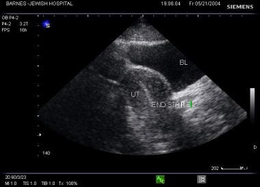 Sagittal viewing showing bladder, uterus (behind the bladder), and endometrial stripe (within the uterus).
When scanning in the sagittal plane, assess for free fluid in the cul-de-sac, also known as the rectouterine pouch or the pouch of Douglas. A small amount of hypoechoic free fluid can be considered physiologic. Fluid tracking two thirds of the way up the posterior wall of the uterus is regarded as moderate. A larger amount of fluid, or fluid that tracks anteriorly and interposes between the uterus and the bladder, is considered large. [32]
Apply acoustic gel to the patient’s right upper quadrant, scanning in the transverse and sagittal planes again to visualize the Morison pouch. Free fluid is generally not visualized until at least 500 mL of free fluid has accumulated; such accumulation is highly suggestive of ruptured ectopic pregnancy (see the image below). [31]
Transvaginal ultrasonography
Transvaginal ultrasonography is best performed on a patient who has an empty bladder.
Position the patient in the supine lithotomy position, preferably on a stretcher equipped for pelvic exams (ie, with stirrups). Scan the uterus in both long and short axial planes. [33]
The cul-de-sac is formed by the peritoneal reflection anterior and posterior to the uterus. A small amount of anechoic fluid in the cul-de-sac is physiologic. Echogenic fluid in the cul-de-sac is highly suggestive of a ruptured ectopic pregnancy.
Upon visualization of a round anechoic structure in the endometrial cavity that is consistent with a gestational sac, acquire pictures and measure the length, height, and width of the gestational sac to obtain the mean sac diameter (MSD). These measurements are taken from the inner aspects of the echogenic border of the sac.
Visualization of a double decidual ring (ie, 2 echogenic rings around the gestational sac) is pathognomonic for an early IUP. In radiology literature, this is considered the earliest reliable sign of an IUP. Double decidual signs, however, are not consistently seen, and caution should be used in terms of using them to determine the presence or absence of an IUP. Prior to this stage, demonstration of a simple gestational sac is an indeterminate ultrasonographic finding.
The yolk sac is a round echogenic ring with an anechoic center located within the gestational sac. The yolk sac can generally be used to determine the presence or absence of an IUP.
The embryo is measured using crown-rump length, which is measured end-to-end (not including the yolk sac).
An extraovarian adnexal echogenic ring (tubal ring sign), highly suggestive of an ectopic pregnancy, occurs when the fallopian tube develops a trophoblastic reaction to an ectopic gestational sac.
Interstitial ectopic pregnancy is rare but has a higher mortality rate following rupture, as the area is richly vascular. An eccentrically located gestational sac with a thin or incomplete myometrial mantle around the sac is suggestive of an interstitial gestation; this is the interstitial line sign.
-
Transverse probe placement for transabdominal obstetric examination.
-
Sagittal or longitudinal probe placement for transabdominal obstetric examination.
-
Sagittal viewing showing bladder, uterus (behind the bladder), and endometrial stripe (within the uterus).
-
Picture of gestational sac with yolk sac.
-
Transverse picture of intrauterine pregnancy.
-
Transverse picture of gestational sac with yolk sac.
-
Picture of abnormal endometrium in a patient with an ectopic pregnancy.
-
Picture of uterus without a fetal pole and a complex adnexal mass consistent with ectopic pregnancy.
-
Free fluid in the Morison pouch from a ruptured ectopic pregnancy.
Tables
| Sonographic Finding | Gestational Age (weeks) Identified by Transvaginal Ultrasound | Gestational Age (weeks) Identified by Transabdominal Ultrasound |
|---|---|---|
| Gestational Sac | 4.5 - 5 | Unable to visualize |
| Yolk Sac | 5 - 6 | 6 - 7 |
| Fetal Pole | 6 | 7 |


