- 2
- ,
- 3
- 8
- 1
To Quiz Yourself: Select OFF by clicking the button to hide the diagnosis & additional resources under the case.
Quick Browser: Select ON by clicking the button to hide the additional resources for faster case review.
CASE NUMBER
372


Diagnosis
Anaplastic Oligodendroglioma
Note
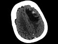
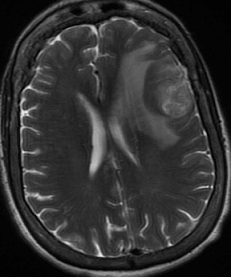
There is an avidly enhancing heterogeneous mass centered in the cortex and subcortical white matter of the left middle and inferior frontal gyri with moderate perilesional edema resulting in mild effacement of the left lateral ventricle and mild rightward midline shift. The CT shows that there is a small area of calcification. The differential diagnosis includes metastasis, oligodendroglioma, and high grade astrocytoma. For a partially calcified frontal lobe mass involving the cortex and subcortical white matter, the best diagnosis is oligodendroglioma which was confirmed on pathology. Enhancement is more common in anaplastic oligodendrogliomas which have a poorer prognosis as was seen in this case.
Related videos to the case