

Case Report
Aplasia Cutis Congenita Mimicking as Scalp Birth Injury in a Neonate: Case Report
1Senior Resident, Department of obstetrics and gynecology, SMS Medical College Jaipur, Rajasthan, India.
2Assistant Professor, Department of Pediatrics, SMS Medical College Jaipur, Rajasthan, India.
3Associate Professor, Department of Pediatrics, SMS Medical College Jaipur, Rajasthan, India.
*Corresponding Author: Sunil Gothwal, Assistant Professor, Department of Pediatrics, SMS Medical College Jaipur, Rajasthan, India.
Citation: Yadav V, Gothwal S, Choudhary R. (2023). Aplasia Cutis Congenita Mimicking as Scalp Birth Injury in a Neonate: Case Report, Journal of Women Health Care and Gynecology, BioRes Scientia publishers. 2(2):1-3. DOI: 10.59657/2993-0871.brs.23.010
Copyright: © 2023 Sunil Gothwal, this is an open-access article distributed under the terms of the Creative Commons Attribution License, which permits unrestricted use, distribution, and reproduction in any medium, provided the original author and source are credited.
Received: July 28, 2023 | Accepted: August 19, 2023 | Published: August 24, 2023
Abstract
Aplasia cutis congenita (ACC) is a rare congenital skin defect. Exact aetiology of ACC is not well understood. Here, we report a case of aplaia cutis congenita mimicking as scalp injury at birth.
Keywords: aplasia cutis congenita; scalp; birth injury
Introduction
Aplasia cutis congenita (ACC) is a rare malformation. It occurs at the scalp, trunk and limbs, and in isolation or as part of group of syndromes [1]. It is a focal defect of cutaneous tissues ranging from an absence of skin to full thickness with bone and dura. Scalp lesions may associate with complications including infection, hemorrhage, thrombosis and seizures. Etiology of ACC is multi-factorial and genetics and exogenous factors possibly play role. Exogenous causes are placental infarcts, teratogens, intrauterine infections, trauma and neural tube defects. Lesions are self-limiting, but warns a workup to screen for underlying soft tissue anomalies [2].
Birth trauma is an injury to the newborn sustained during the process of birth with an incidence of 2-7 per 1000 live births [3]. It may vary from superficial injury of skin to life threatening problems [4]. In our case the newborn baby had congenital localized absence of skin over the centre of scalp region with well-defined margins. Initially it looked like as an avulsion injury by artery forceps during LSCS. It may cause an apprehension to the obstetrician, as the obstetricians are often not aware of this rare congenital skin defect.
Case Report
A 29 years old female, G3P1L1A1 with previous lower segment cesarean section (LSCS) delivered, at term gestation at a tertiary care centre in Rajasthan, India by LSCS and gave birth to a 2.5 kg male baby. Antenatal period was uneventful. Though, history of anti-tubercular therapy before she conceived and of beta blockers during 3rd trimester for hypertension was there. We noticed that the newborn had a single skin lesion of about 2X2 cm diameter, oval in shape with well-defined margins involving epidermis and dermis on the scalp in the midline at lambda point of scalp (Figure 1). At first the lesion was wrongly taken as instrumental birth injury. The newborn had normal agar scores n vitals were stable. Later, clinical diagnosis of aplasia cutis congenita was made and the baby was admitted in NICU for further evaluation. There was no history of ACC causing drugs during pregnancy. As mother of our case took ATT and beta blocker, we did not find any association of these with ACC in available literature. Other associated anomalies were ruled out as ultrasonography scan was normal. As the lesion was small, less than 4 cm, local cleansing with topical anti-bacterial ointment was done and the baby was put on regular dermatology OPD follow up. Later at 6 weeks of age the lesion was completely filled without any local complication.
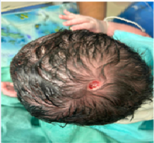
Figure 1: Single skin lesion of about 2X2 cm diameter, oval in shape with well-defined margins involving epidermis and dermis on the scalp in the midline at lambda point of scalp.
Discussion
Aplasia cutis congenita (ACC) is an uncommon anomaly of newborns with an incidence of approximately 1 to 3 out of 10,000 births [5]. Gender predilection is not reported. It manifests as a solitary defect on the scalp in 70% of cases, but it may also occur as multiple lesions. Although commonly seen on the scalp yet it can affect any part of the body. Lesions are non-inflammatory and well-demarcated with variable size. They may be circular, oval and linear in shape. At birth, the lesions may have healed, may superficially erode and may deeply ulcerate. Defects in the skin that form early in gestation may heal before delivery and appear as an atrophic, membranous, bullous, or parchment like scar with associated alopecia, whereas less mature defects present as ulcerations. The membranous type of aplasia cutis congenita is most common.
The exact pathophysiology of ACC is unclear. The most commonly accepted theory focuses on the tension that prevents the skin from converging during development in utero. Proposed mechanisms include intrauterine trauma, vascular compromise, infection, and medications. Known medicines are methimazole, misoprostol and valproic acid. Aplasia cutis congenita is mostly sporadic, though autosomal dominant and recessive cases have also been reported. Mutations in the ribosomal GTPase BMS1 have been identified.
Newborns with ACC need to be thoroughly examined for associated anomalies including bone and intracranial malformations. Ultrasound imaging can be used to evaluate for underlying defects [6]. Further tests may be indicated for evaluation of associated conditions. Differential diagnoses are traumatic lesions, localized scalp infections, dermoid cyst, isolated encephalocele, meningocele and nodular neuronal heterotopia.
ACC management in neonates is based on the size of skin defect. For small lesions (<4cm>4cm) are more commonly associated with underlying defects and are at an increased risk of complications, including hemorrhage, venous thrombosis, and infection. Early surgical repair is recommended to avoid these complications. Skin grafting or flap techniques are commonly utilized as some lesions can be several centimeters in size.
The prognosis for aplasia cutis congenita (ACC) is usually excellent for small skin defects. The recovery is uneventful, with gradual epithelialization and formation of a hairless, atrophic scar over several weeks. The average length of recovery for patients treated conservatively is 27.9 days [8]. Small underlying bony defects usually close spontaneously during the first year of life. Surgical repair of large or multiple scalp defects with excision and primary closure may be considered. If aplasia cutis congenita is associated with other anomalies, the prognosis is dependent on the severity of the associated abnormalities.
Conclusion
This case report retraites the importance of antenatal history, careful examination of skin lesion, and pediatrician opinion before labelling it as birth injury.
References
- Verhelle NA, Heymans O, Deleuze JP, Fabre G, Vranckx JJ, Van den hof B, et al. (2004). Abdominal aplasia cutis congenita: case report and review of the literature. J Pediatr Surg, 39:237-239.
Publisher | Google Scholor - Humphrey SR, Hu X, Adamson K, Schaus A, Jensen JN, Drolet B. (2018). A practical approach to the evaluation and treatment of an infant with aplasia cutis congenita. J Perinatol, 38(2):110-117.
Publisher | Google Scholor - Pressler JL. (2008). Classification of major newborn birth injuries. J Perinat Neonatal Nurs, 22:60-67
Publisher | Google Scholor - Mazza F, Kitchens J, Akin M, Elliott B, Fowler D, Henry E, et al. (2008). The road to zero preventable birth injuries. JtComm J Qual Patient Saf, 34:201-205.
Publisher | Google Scholor - E Rogvi R, Sommerlund M, Vestergaard ET. (2014). Aplasia cutis congenita is a rare and possibly overlooked congenital anomaly. Ugeskr Laeger, 24:176(48).
Publisher | Google Scholor - Browning JC. (2013). Aplasia cutis congenita: approach to evaluation and management. Dermatol Ther, 26(6):439-444.
Publisher | Google Scholor - Belkhou A, Francois C, Bennis Y, Duquennoy-Martinot V, Guerreschi P. (2016). Aplasia cutis congenita: Update and management. Ann ChirPlastEsthet, 61(5):450-461.
Publisher | Google Scholor - Tempark T, Shwayder TA. (2012). Aplasia cutis congenita with fetus papyraceus: report and review of the literature. Int J Dermatol, 51(12):1419-1426.
Publisher | Google Scholor


