
Lampyris noctiluca ( Linnaeus, 1758 )
|
publication ID |
https://doi.org/ 10.11646/zootaxa.4247.4.5 |
|
publication LSID |
lsid:zoobank.org:pub:92D9E2D4-5BA0-4446-BB98-267E231A4737 |
|
DOI |
https://doi.org/10.5281/zenodo.6020808 |
|
persistent identifier |
https://treatment.plazi.org/id/3139F14C-6123-AF19-0BAC-FF2FFE3B338F |
|
treatment provided by |
Plazi |
|
scientific name |
Lampyris noctiluca ( Linnaeus, 1758 ) |
| status |
|
Lampyris noctiluca ( Linnaeus, 1758) View in CoL
Material examined. Ljubljana ( Slovenia), three higher instar larvae out of six collected in the first half of September 2013 and 2015, three pupae reared from remaining collected larvae.
Diagnosis. Larvae robust, dark brown or black; with light coloured spots on posterolateral margins on pronotum and every tergite except caudal segment; pronotum concave on posterior margin; maxillary palpomere II with inner-lateral sagittal slot; mandibles with retinaculum forming a sharp inner tooth; mandibular channel covered with thick blunt seta; microscopic granulose protuberances, densely occurring on sclerites and legs; photic organ represented by a pair of conspicuous whitish spots located on pleurites of abdominal segment VIII.
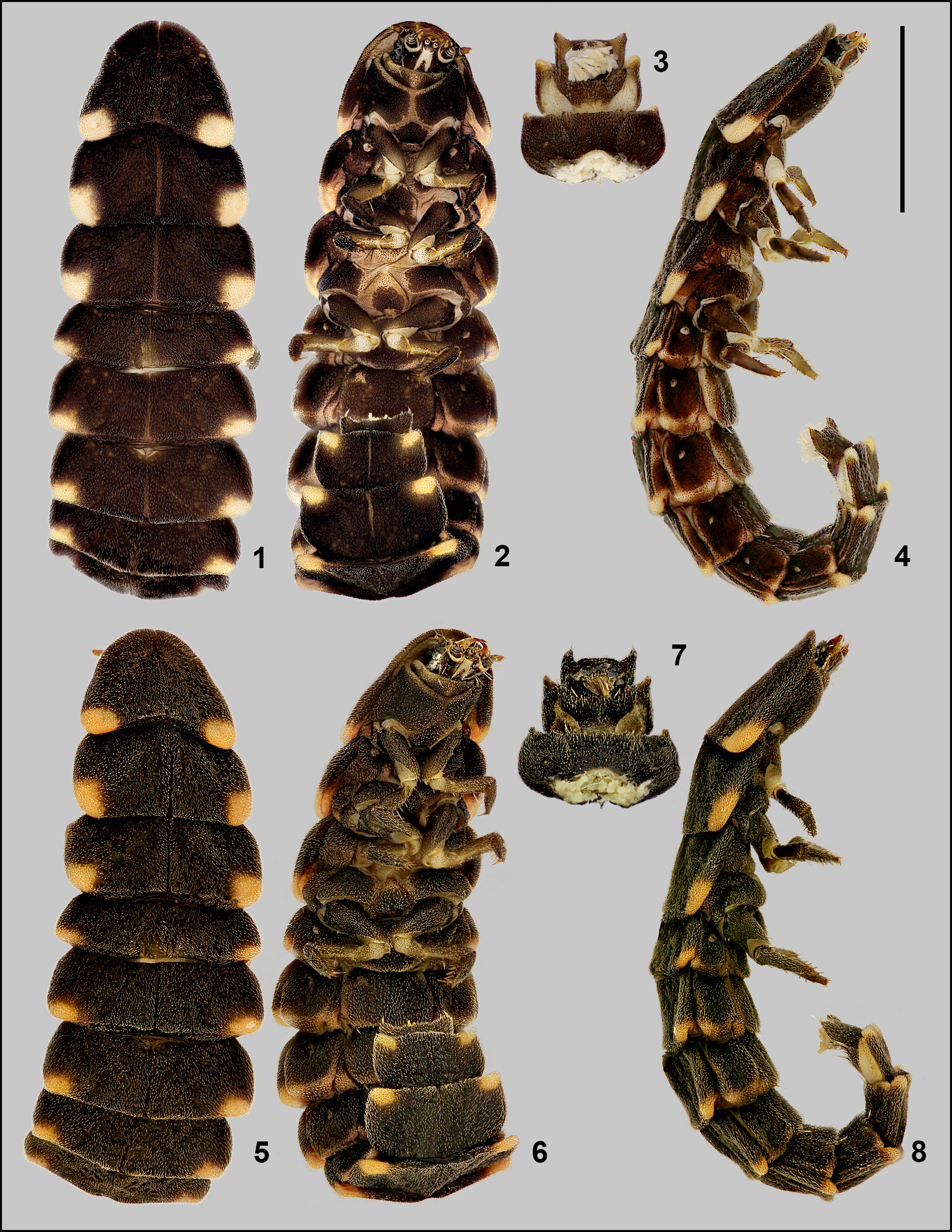
Description of mature larva ( Figs 1–8 View FIGURES 1 – 8 ). Fusiform and robust; slightly flattened dorsoventrally. Body length 20–23 mm (from the anterior margin of pronotum to the apex of caudal segment); with 3 thoracic and 10 abdominal segments. Tergites from pronotum to abdominal segment IX extending laterally to cover the rest of the body and divided by sagittal line in dorsal view. Colouration: body dark brown or black, with distinct pinkish or yellowish spots on posterolateral margins on pronotum and every tergite except caudal segment. Spiracles on pleural plates of light colouration. Photic organ represented by a pair of whitish spots located on pleurites of abdominal segment VIII.
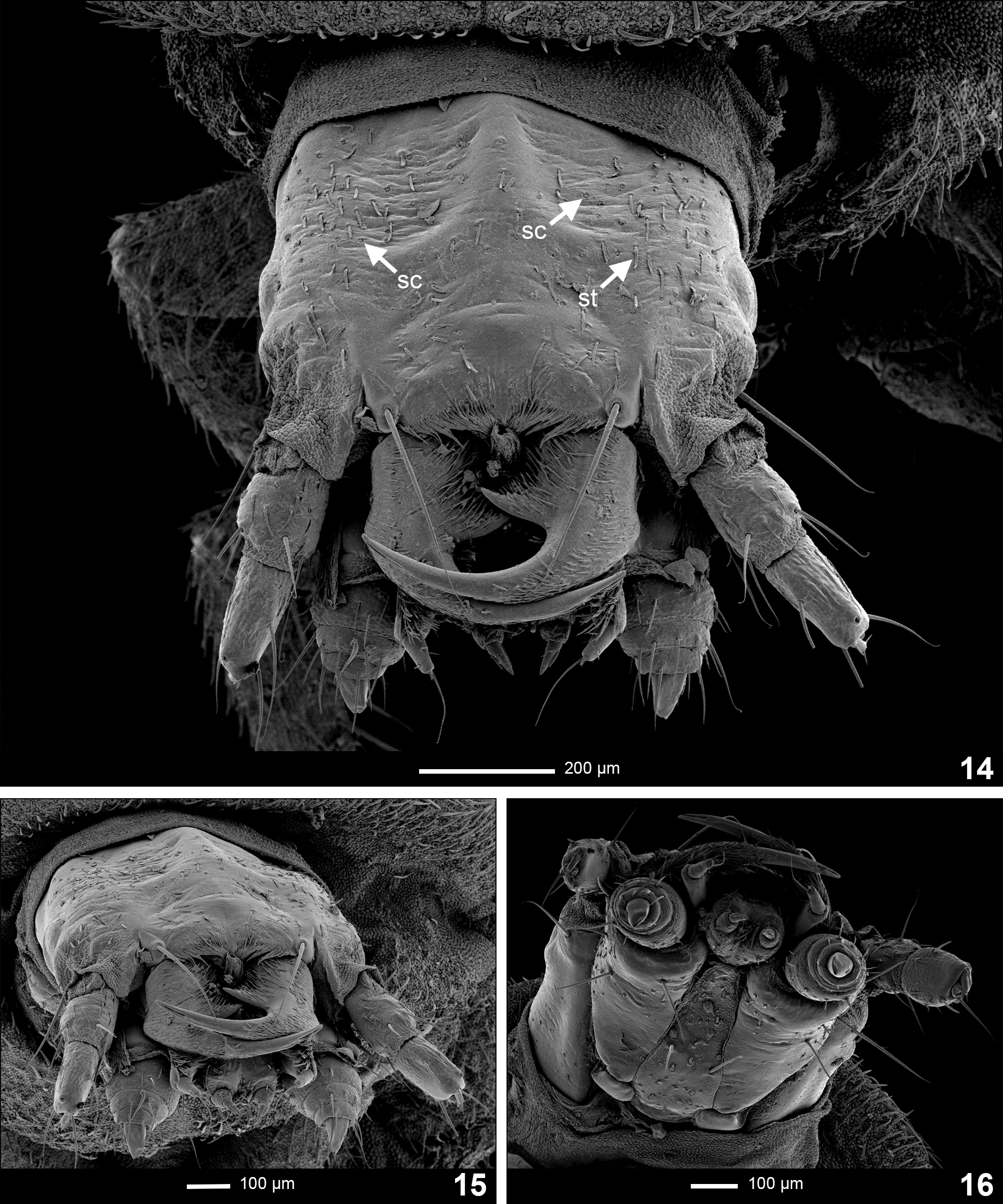
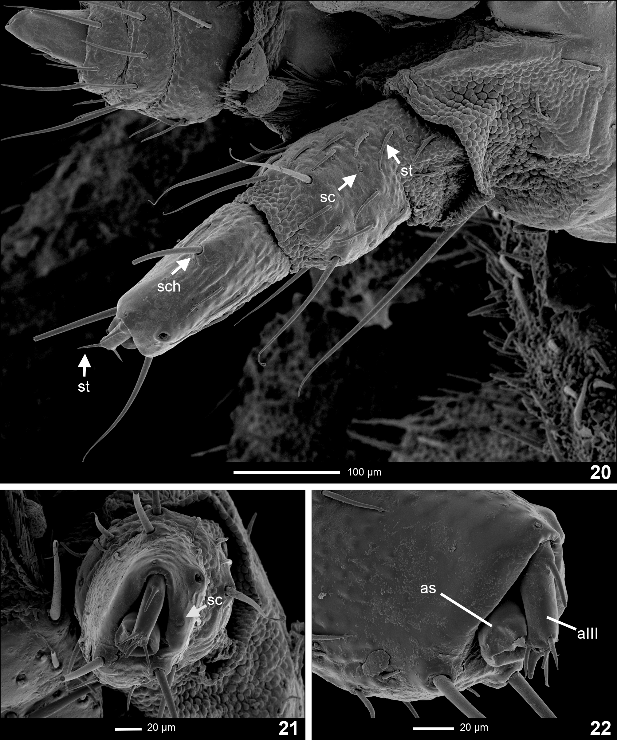
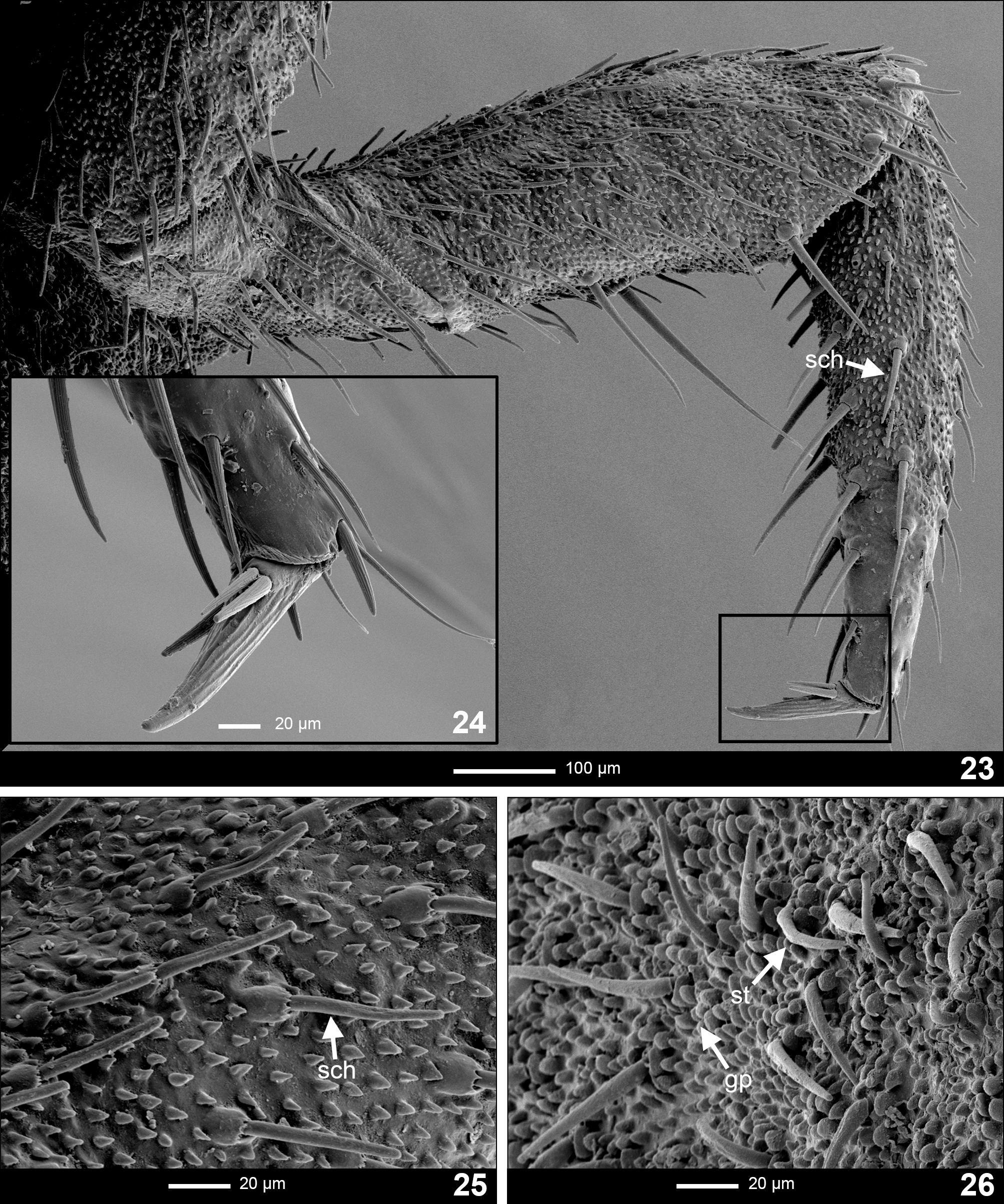
Cuticular outgrowths). 1. Stout, short, blunt, oblique setae ( Figs 14 View FIGURES 14 – 16 , 20 View FIGURES 20 – 22 , 26 View FIGURES 23 – 26 ; st); 2. dense granulose protuberances ( Fig. 26 View FIGURES 23 – 26 ; gp); 3. long stout setae ( Figs 20 View FIGURES 20 – 22 , 23, 25 View FIGURES 23 – 26 ; sch); 4. coeloconical receptors ( Figs 14 View FIGURES 14 – 16 , 20, 21 View FIGURES 20 – 22 ; sc).
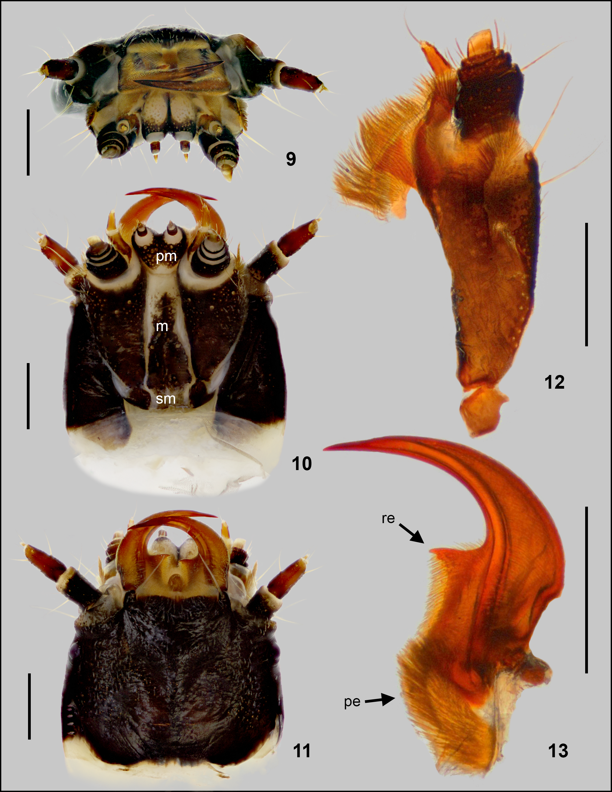
Head capsule ( Figs 9 – 11 View FIGURES 9 – 13 , 14–16 View FIGURES 14 – 16 ). Prognathous; retractable within prothorax, extensible neck membrane forming a two layered envelope around retracted head; of equal width and length; slightly widening posteriorly. Epicranial plate laterally about the same size as width of head capsule at its shortest width, with one stout seta anterolateraly close to the base of antenna. Head capsule dorsally covered with short blunt setae lying on surface and coeloconical receptors ( Figs 14 View FIGURES 14 – 16 , 21 View FIGURES 20 – 22 ; sc). Epicranial suture hardly distinguishable for its dark pigmentation, but present ( Fig. 11 View FIGURES 9 – 13 ). One stemma on each side of head. Labrum fused to clypeus forming a clypeolabrum, covering base of mandibles in dorsal view. Clypeolabrum slightly double-arched in anterior view, with one long seta on each lateral corner, reaching the apex of mandibles ( Fig. 14 View FIGURES 14 – 16 ). Epipharynx formed by two plates, and an anterior pair of brushes of long setae on each plate, which project centrally past anterior margin of head. Hypopharynx covered with long setation. Gula absent ( Fig. 10 View FIGURES 9 – 13 ).
Antenna ( Figs 20–22 View FIGURES 20 – 22 ). Trimerous, inserted on lateral distal margin of epicranial plate; partially retractable within membranous socket. Basal antennomere widest, fully sclerotized, bearing short setae lying on surface, coeloconical receptors and several long oblique setae near the apical region ( Fig. 20 View FIGURES 20 – 22 ). Several long stout setae placed radially on the anterior margin, with a distinct seta on inner lateral area of antennomere. Second antennomere slightly shorter than first, laterally flattened; bearing numerous short setae lying on surface irregularly scattered over the antennomere, together with several coeloconical receptors ( Fig. 21 View FIGURES 20 – 22 ; sc) and four stout setaefirst three in the middle and on apex of inner margin of antennomere, fourth on apical region of outer lateral margin of antennomere. Sensorium of second antennomere ( Figs 21, 22 View FIGURES 20 – 22 ; as) oval, widest at the base, closely touching second antennomere, shorter than third antennomere with no visible surface pattern. Third antennomere ( Figs 21, 22 View FIGURES 20 – 22 ; aIII) shortest, bearing four short setae; one basal and three apical, together with a pair of short cuticular projections; ventrally apical thick and dorsal-apical thin.
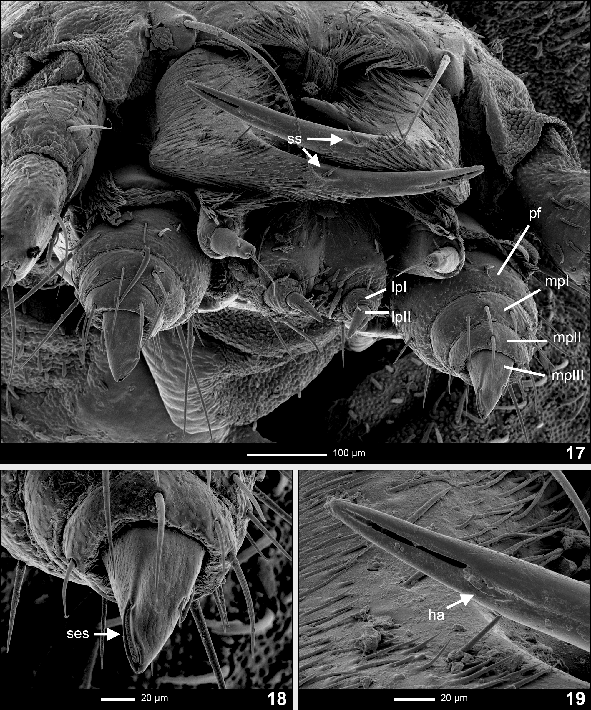
Maxilla ( Fig. 12 View FIGURES 9 – 13 ). Consisting of five parts, attached by membrane to labium forming a maxillo-labial complex ( Fig. 17 View FIGURES 17 – 19 ). Cardo transverse, sub-rectangular, slightly wider than long. Stipes elongated, ventrally relatively glabrous, setae mainly on distal half, with three long stout setae placed radially on ventral apical region; dorsolaterally covered with short setation lying on surface. Galea bimerous, basal part sub-cylindrical, slightly wider than distal, with long dorsal setation partially covering distal part; distal part sub-cylindrical, inclined centrally, with short setae and one apical seta longer than body of the distal part. Lacinia with a brush of long setae on outer lateral margin. Palpifer ( Fig. 17 View FIGURES 17 – 19 ; pf) large, rectangular, about the same length and width. Maxillary palpus trimerous ( Figs 17, 18 View FIGURES 17 – 19 ; mpI, mpII, mpIII), basal and second palpomere short and wide. Palpifer and palpomeres I– II covered with several setae mainly on outer dorsolateral margin; palpomere III ( Fig. 18 View FIGURES 17 – 19 ; mpIII) irregularly subconical, thick, blunt, with an inner longitudinal lateroapical sensory slot ( Fig. 18 View FIGURES 17 – 19 ; ses), small seta on outer lateral region and short outer lateral longitudinal sensory slot covered with thin seta lying on surface.
Labium ( Fig. 17 View FIGURES 17 – 19 ). Closely attached to maxilla, formed by a short and strongly sclerotized prementum, mentum and weakly sclerotized submentum ( Fig. 10 View FIGURES 9 – 13 ; pm, m, sm). Glossae absent. Prementum heart-shaped in ventral view; covered with very short setation; bearing several longer blunt setae, and a pair of long stout setae, placed centrally on ventral region. Labial palpus bimerous ( Fig. 17 View FIGURES 17 – 19 ; lp1, lp2); basal palpomere wider than long, bearing several setae; distal palpomere conical, longer and narrower than basal, bearing a short thin erect seta on basal half dorsally, a longer, stout and blunt seta covering a sagittal slot positioned on outer margin and sensillum coeloconicum on outer ventrolateral side of apex. Mentum elongated, sub-triangular, unsclerotized on lateral margins, ventrally bearing numerous short setae lying on surface and a pair of long, erect setae centrally.
Mandible ( Figs 13 View FIGURES 9 – 13 , 19 View FIGURES 17 – 19 ). Symmetrical, falcate, with an internal channel opening subapically on outer edge ( Fig. 19 View FIGURES 17 – 19 ). Penicillus well developed ( Fig. 13 View FIGURES 9 – 13 ; pe). Retinaculum present, forming one sharp inner tooth on basal half of mandible ( Fig. 13 View FIGURES 9 – 13 ; re). Inner margin of mandible from retinaculum to the base covered with stout setae ( Fig. 13 View FIGURES 9 – 13 ). Basal two-thirds of mandible ventrally with dense setation lying on surface and aimed centrally. Dorsally, mandible covered with several strong setae lying on surface, aiming medially on proximal two-thirds of each mandible. Lateral margin covered by brush of short setae lying on surface of basal two-thirds ( Fig. 17 View FIGURES 17 – 19 ). Sensory (hyaline) appendage on outer margin of mandible before channel opening consists of a blunt thick seta ( Fig. 19 View FIGURES 17 – 19 ; ha). A distinct short, stout seta set in a shallow depression present dorsally at anterior end of lateral setation ( Fig. 17 View FIGURES 17 – 19 ; ss).
Thorax ( Figs 1, 2, 4–6, 8 View FIGURES 1 – 8 ). Three-segmented, thoracic tergites divided by sagittal line ( Figs 1, 5 View FIGURES 1 – 8 ). Pronotum of equal length and width, sub-trapezoidal, wider posteriorly, rounded at posterolateral corners, strongly concave on posterior margin. Meso- and metanotum sub-rectangular, wider than long, mesonotum longer than metanotum. Lateral areas of meso- and metathorax formed by episternum and epimeron; episternum of mesothorax bearing an annular spiracle. Prosternum rounded, wider than long, robust, well sclerotized, subdivided into three plates; lateral ones extending above and to the sides of coxae; medial plate sub-pentagonal. Meso- and metasternum subdivided by transverse fold into poorly sclerotized basisternum and well sclerotized sternellum; sternellum subdivided into three plates, lateral ones extending above and to the sides coxae, representing large episterna and smaller epimera, medial plate less sclerotized on margins, heart-shaped with wider margin posteriorly.
Legs ( Figs 23, 24 View FIGURES 23 – 26 ). Pentamerous, all pairs similar in shape and size. Coxa large, stout, covered by short sharp setae. Coxal-trochanteal membrane ( Figs 2, 6 View FIGURES 1 – 8 ) reaching less than 1/2 of coxal length. Trochanter small, subtriangular in lateral view, shorter than femur, covered by short sharp setae. Femur slightly fusiform, widening towards apex in lateral view, covered by short sharp setae, with several large setae ventrally. Tibiotarsus as long as femur, narrower, tapering towards distal end, bearing stout short sharp setae dorsally and strong sharp erect setae ventrally. Pretarsus ( Fig. 24 View FIGURES 23 – 26 ) composed of a claw with distinct ridges, ventrally bearing three short stout setae with fine ridges. Cuticle of leg ( Fig. 25 View FIGURES 23 – 26 ) densely covered with grainy protuberances except for apical half of tibiotarsus ( Fig. 23 View FIGURES 23 – 26 ).
Abdomen ( Figs 1–8 View FIGURES 1 – 8 ). Ten-segmented, tapering towards posterior end, segments I to VIII subdivided by fine sagittal line in dorsal view. Tergites of segments I to VIII sub-trapezoidal, similar in shape and colouration, wider than long; tergite of segment IX sub-rectangular; segment X forming a narrow, incompletely sclerotized dark ring, bearing the holdfast organ—pygopod ( Fig. 3 View FIGURES 1 – 8 )—with several eversible processes. Ventrites of segments I to VIII sub-rectangular, slightly wider than long, well sclerotized, with a pair of long stout setae on posterolateral margins; ventrite of segment IX sub-trapezoidal. Pleural areas well sclerotized, pleural suture of segments I to V subdivide lateral areas into large sub-rectangular upper pleurite, bearing an annular spiracle, and narrow lower pleurite anteriorly covered; pleural segments VI to VIII with only upper pleurite bearing an annular spiracle. Segment VIII bearing photic organs ventrally on pleurites, appearing as two whitish spots ( Fig. 3 View FIGURES 1 – 8 ).
Larval behaviour. Larvae of Lampyris noctiluca were observed to “ride” the snails, which means mounting the shell and assuming a favourable position to inject the head of the snail or its upper tentacles with lethal toxin. The larva then remains on the shell until the snail shows no signs of life. Feeding can take up to two days. The larva avoids feeding on digestive organs and can sometimes remove these organs from the shell before resuming feeding on the rest of the body. After feeding, the larva remains passive for certain time and after ca. one day excretes a dark brown fluid from the back of its abdomen.
While collecting larvae in nature, a several seconds lasting glow was often observed, followed by a long period of darkness. In captivity, the larvae demonstrated this behaviour when disturbed. When undisturbed, the larvae occasionally showed photic behaviour of definite pulses of light lasting ca. 2 seconds, separated from the next one by a longer interval of darkness lasting ca. 4 seconds. The other photic manifestation consisted of continuous glow of weaker intensity, which could take several minutes.
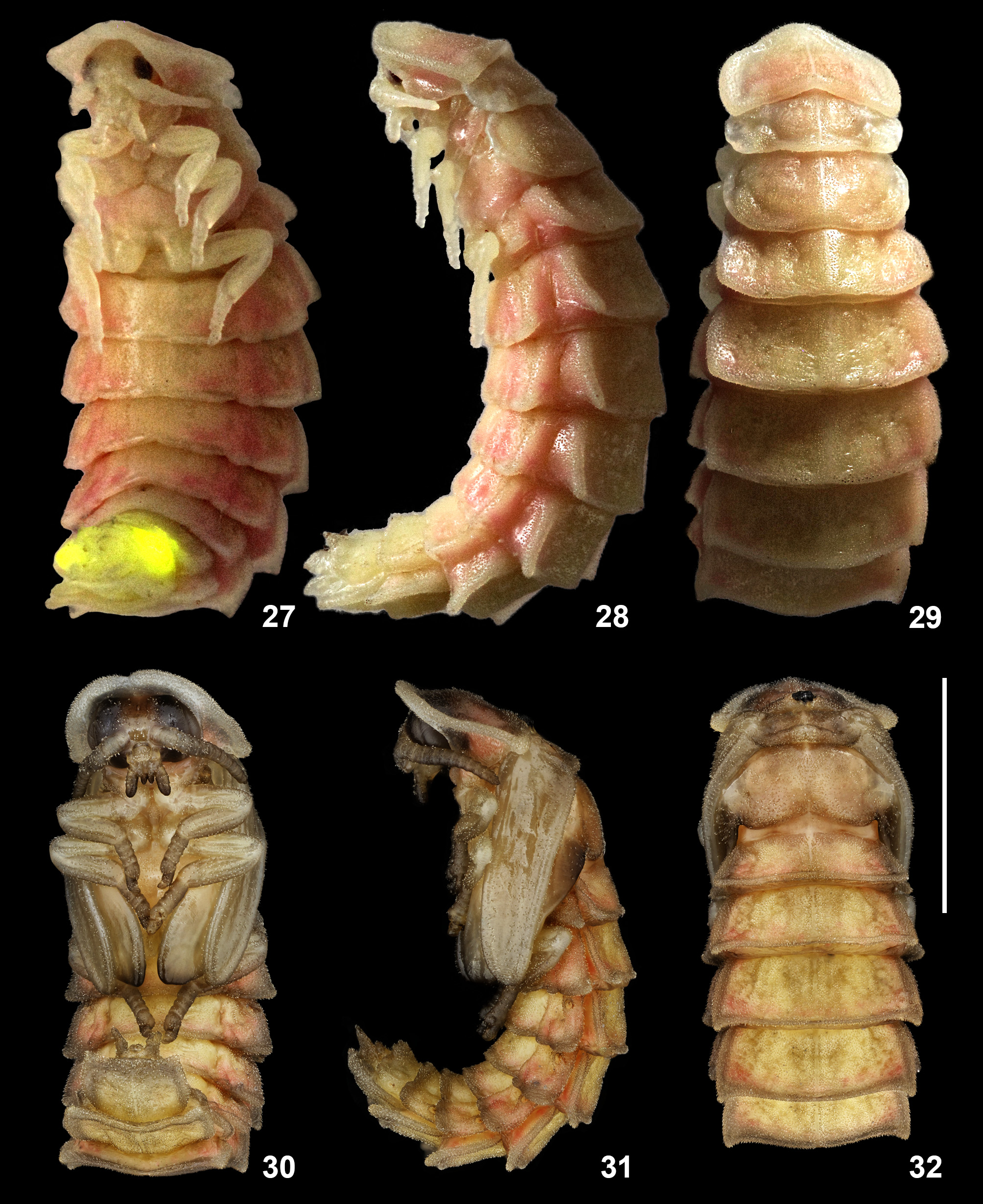
Description of female pupa ( Figs 27–29 View FIGURES 27 – 32 ). Type of pupa: adectica exarata libera. Curved, ventrally concave. Length 20 and 23 mm. Colouration: yellowish white on tergites and parts that will become sclerotized in adult stage, pink especially in pleural region. Surface covered by short setae.
Head capsule. Completely covered by pronotum in dorsal view. Eyes small. Antennae short, extending laterally, without reaching posterolateral corners of pronotum. Mouthparts visible in ventral view.
Thorax. Pronotum similar in shape to that of larva, with more acute posterolateral corners and less elongated. Meso- and metanotum smaller, sub-rectangular; mesonotum significantly shorter than metanotum, bearing very short elytra which become vestigial in adult individual. All pairs of legs free, visible in ventral view, proportionally shorter to the overall body size than those of males. Spiracles present on pleural areas of mesothorax.
Abdomen. Abdominal segments sub-rectangular, wider than long. Ventrites on segments II–IV bearing a double sagittal line of depression in cuticle. Spiracles present on abdominal pleural areas of segments I–VIII. Segment VIII bearing functional larval photic organs ( Fig. 27 View FIGURES 27 – 32 ).
Description of male pupa ( Figs 30–32 View FIGURES 27 – 32 ). Type of pupa: adectica exarata libera. Curved, ventrally concave. Length 17 mm. Colouration on young pupae: ochred yellow on tergites and parts that will become sclerotized in adult stage, pink especially in pleural region in young pupae. Colouration on older pupae: beige to brown with a tint of olive or army green on pronotum, head, elytra and legs; inner surface of dorsal and abdominal tergites ochred yellow. Surface covered by short setae.
Head capsule. Completely covered by pronotum in dorsal view. Eyes distinctly large, on sides of the head. Antennae short, extending laterally towards distal end of prothoracic femur. Mouthparts visible in ventral view.
Thorax. Pronotum semicircular and proportionally longer than that of female when compared to overall body size (length of pronotum to body length ratio is 0.23 while in female the ratio is 0.15). Small narrow mesonotum and large wide metanotum sub-rectangular, bearing beige wing pads with dark brown apices, covered by beige elytra of about the same length; wing pads reaching distal end of second abdominal segment, when pupa is relaxed. Pro- and mesothoracic legs free, visible in ventral view; metathoracic legs almost completely covered by wing pads except for distal segments of tarsi, which extend past second ventrite. Spiracles present on pleural areas of mesothorax.
Abdomen. Abdominal segments sub-rectangular, wider than long. Spiracles present on abdominal pleural areas of segments I–VIII. Segment VIII bearing functioning larval photic organs.
Pupal behaviour. Both male and female pupae are commonly idle, either lying on their side or back, responding only to disturbance. A luminescent response can be induced by handling the animal or even by vibrations. It consist of one short glow, lasting several seconds, with a peak intensity lasting ca. 1 second and then quickly fading away. When under a strong light source, the pupa starts to move its abdomen, twiddling from side to side or doing “crunches”. This behaviour suggests an effort to move into darker area, i.e. positive thigmotaxis. The same behaviour can be observed in prepupae.
Development period and ontogenetic morphological changes in pupae. From the reared larvae, future male entered the stage of prepupa 1 day prior to pupation and future females 3–6 days prior to pupation. The pupal period lasted 8 days for male and 7–10 days for females.
The mesonotum of a female is laterally blunt in the first few days of pupal development, but later the lateral margins begin to sharpen to form vestigial elytra. On the other hand, in male pupae, the elytra and wings are already semi developed since the first day of the pupal stage. Therefore, the sex of future adult can be determined in any time of the pupal period.
No known copyright restrictions apply. See Agosti, D., Egloff, W., 2009. Taxonomic information exchange and copyright: the Plazi approach. BMC Research Notes 2009, 2:53 for further explanation.